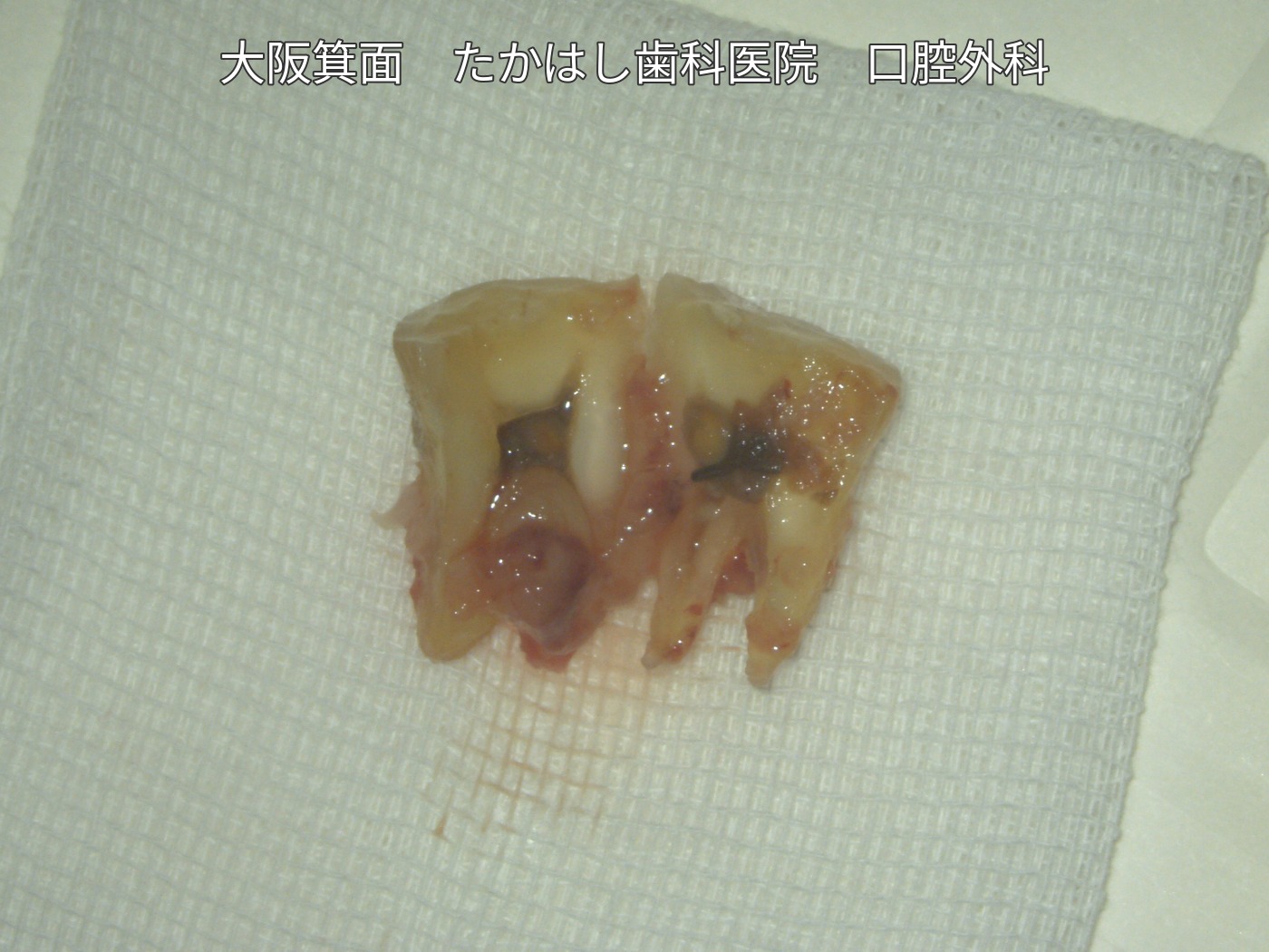
Adhesive repair and replantation of a lower right molar that had split into two pieces
This case involved extraoral adhesive repair and treatment of infected root canals in a fractured lower right first molar, followed by replantation in its original position. Actual footage shows the mobility of the crown, followed by photographs documenting treatment through zirconia crown placement.
Case video
Treatment, cost, duration and number of visits for this case
Treatment: Extraoral adhesive repair and treatment of infected root canals, replantation and temporary splinting, and final restoration with a zirconia crown.
Total cost: JPY 640,000 (tax included, including placement of the final zirconia crown)
Treatment duration: Approximately 3 months
Number of visits: 9 visits
The visits consisted of the initial consultation, an explanation of the treatment, surgery, follow-up at 1, 2, 4 and 8 weeks after surgery, an impression at 12 weeks, and crown placement. Treatment duration and the number of visits vary by case.
Main risks and treatment limitations
Reinfection, root resorption, ankylosis (fusion of the tooth to bone), or poor healing after replantation may occur, and further treatment or extraction may be necessary. Outcomes vary from person to person; preservation of the tooth and its long-term prognosis are not guaranteed. This case record covers the period up to placement of the final crown.
Condition before treatment
The gums around the lower right first molar were swollen, and the crown moved when an instrument was inserted into the fracture. The opening of the video includes actual footage demonstrating this mobility.
Extraoral examination and repair
After removal and examination, the tooth was found to be in two pieces, with dark contamination inside. Adhesive repair and treatment of the infected root canals were performed outside the mouth.
From replantation and temporary splinting to the final restoration
The repaired tooth was returned to its original position and temporarily splinted to the tooth in front of it. At 3 months after surgery, progress was checked and an impression was taken for the crown. The final photograph shows the zirconia crown in place.
The case photographs and video are used with the patient’s consent to publication.