
Tooth-preserving treatment for root fractures in Minoh, Osaka.
- Case 1 | Root fracture and odontogenic maxillary sinusitis in an upper right second molar: intentional replantation
- Case 2 | Root fracture in an upper right first premolar: adhesive repair and intentional replantation
- 01 Gum swelling and pain
- 02 A tooth that looked healthy at first glance
- 03 A crack identified on close examination
- 04 Examining the split from another angle
- 05 Removing the tooth from the mouth for repair
- 06 Examining the tooth outside the mouth
- 07 A tooth completely split in two
- 08 Returning the repaired tooth to its original position
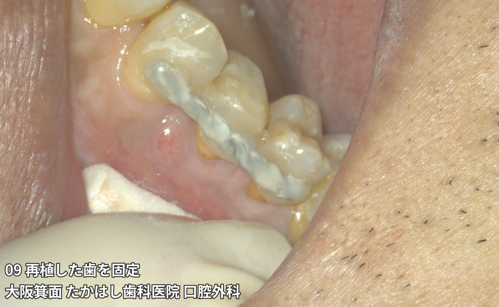
- 09 Splinting the replanted tooth
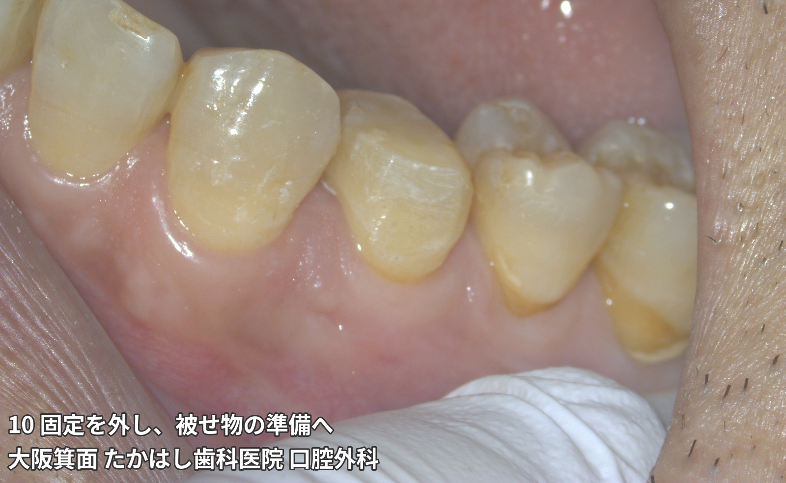
- 10 Removing the splint and preparing for a crown
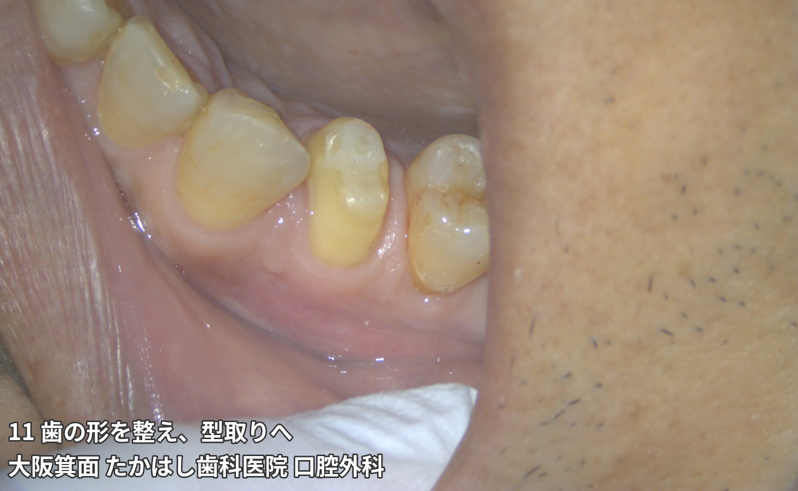
- 11 Shaping the tooth and taking an impression
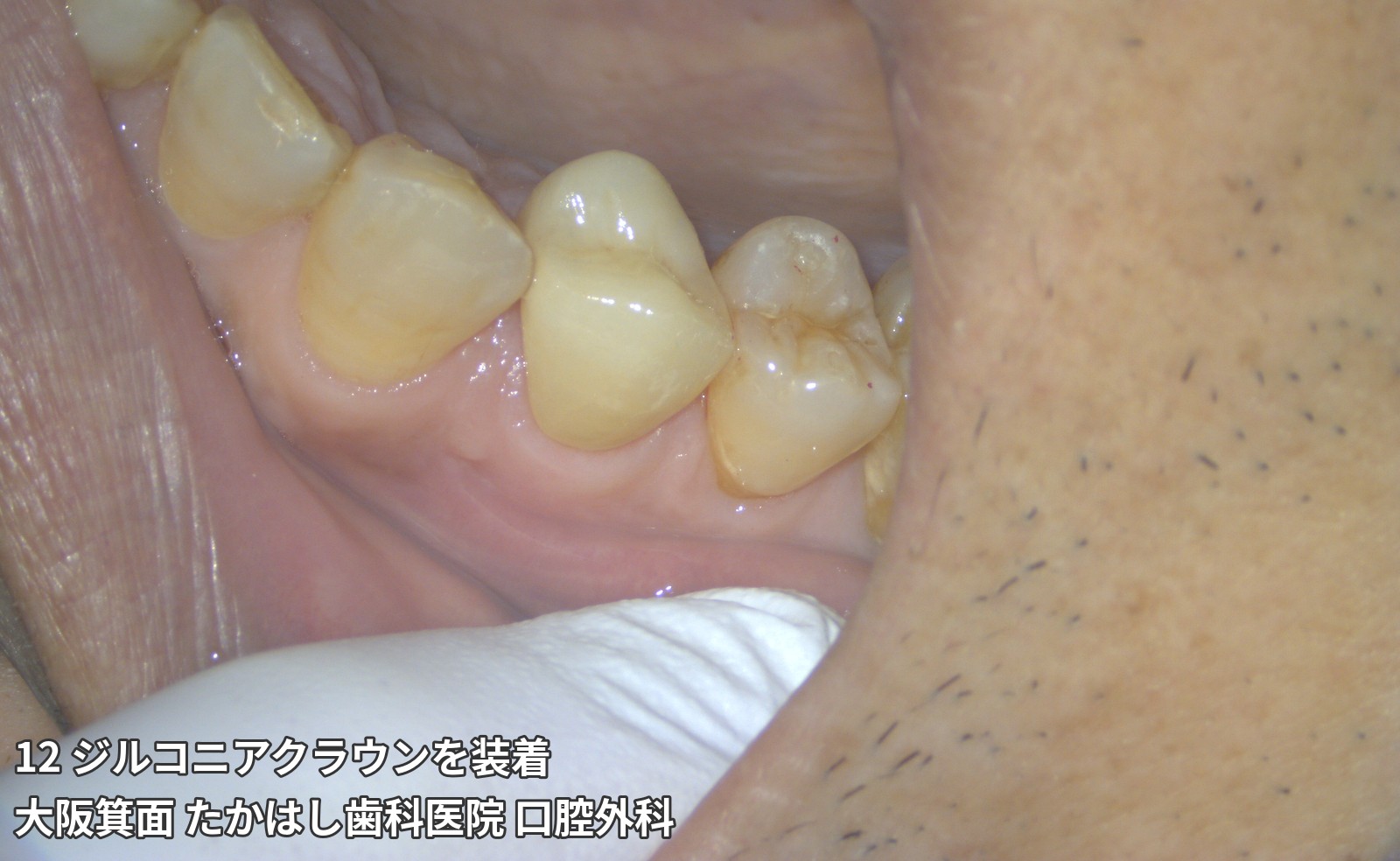
- 12 Placement of a zirconia crown
- What to consider before deciding on extraction
- What is a root fracture? A crack or split in the root of a tooth
- Treatments considered to preserve a tooth
- When preservation is difficult, we explain the reasons and options
- From consultation to follow-up care
- Private treatment: costs, duration, and risks
- Frequently asked questions
- A consultation to help you make an informed choice about a valued tooth
- Case 1 | Root fracture and odontogenic maxillary sinusitis in an upper right second molar: intentional replantation
- Case 2 | Root fracture in an upper right first premolar: adhesive repair and intentional replantation
- 01 Gum swelling and pain
- 02 A tooth that looked healthy at first glance
- 03 A crack identified on close examination
- 04 Examining the split from another angle
- 05 Removing the tooth from the mouth for repair
- 06 Examining the tooth outside the mouth
- 07 A tooth completely split in two
- 08 Returning the repaired tooth to its original position
- 09 Splinting the replanted tooth
- 10 Removing the splint and preparing for a crown
- 11 Shaping the tooth and taking an impression
- 12 Placement of a zirconia crown
- What to consider before deciding on extraction
- What is a root fracture? A crack or split in the root of a tooth
- Treatments considered to preserve a tooth
- When preservation is difficult, we explain the reasons and options
- From consultation to follow-up care
- Private treatment: costs, duration, and risks
- Frequently asked questions
- A consultation to help you make an informed choice about a valued tooth
Case 1 | Root fracture and odontogenic maxillary sinusitis in an upper right second molar: intentional replantation
This patient came to us hoping to save an upper right back tooth that had fractured during a meal. Because the root fracture was accompanied by odontogenic maxillary sinusitis, we performed intentional replantation: removing the tooth, repairing it, and returning it to its original position.
After treatment, improvement in the odontogenic maxillary sinusitis was observed, and the tooth was restored with a zirconia crown. Photographs illustrate the course from the initial consultation through follow-up after treatment.
In this case, treatment took approximately 4 months, involved around 8 visits, and cost JPY 640,000 (tax included; private treatment not covered by health insurance). The course and outcome vary between individuals. This case does not guarantee the same result or long-term tooth retention.
Seeking a way to save the tooth
The patient initially visited another dental clinic after hearing a sound during a meal and finding that a back tooth had fractured. Although extraction was explained, the patient wanted to retain their own tooth if possible and came to our clinic for a consultation. The affected tooth was the upper right second molar (upper right 7). In addition to the fracture, swelling was observed in the surrounding gums.
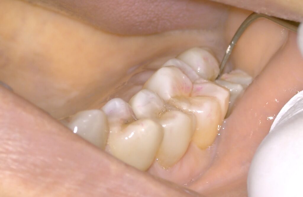
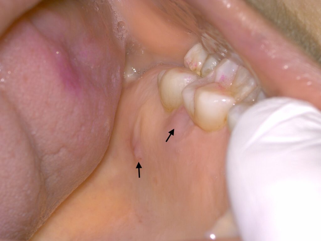
Root fracture and surrounding infection
A vertical fracture extended from the crown to the root of the tooth. Bacteria entering through a fracture can spread infection inside the tooth and into the surrounding tissues.
In this case, odontogenic maxillary sinusitis associated with the dental infection was also present. The maxillary sinus is a cavity near the upper back teeth. Treatment needed to address both the fracture and the surrounding infection.
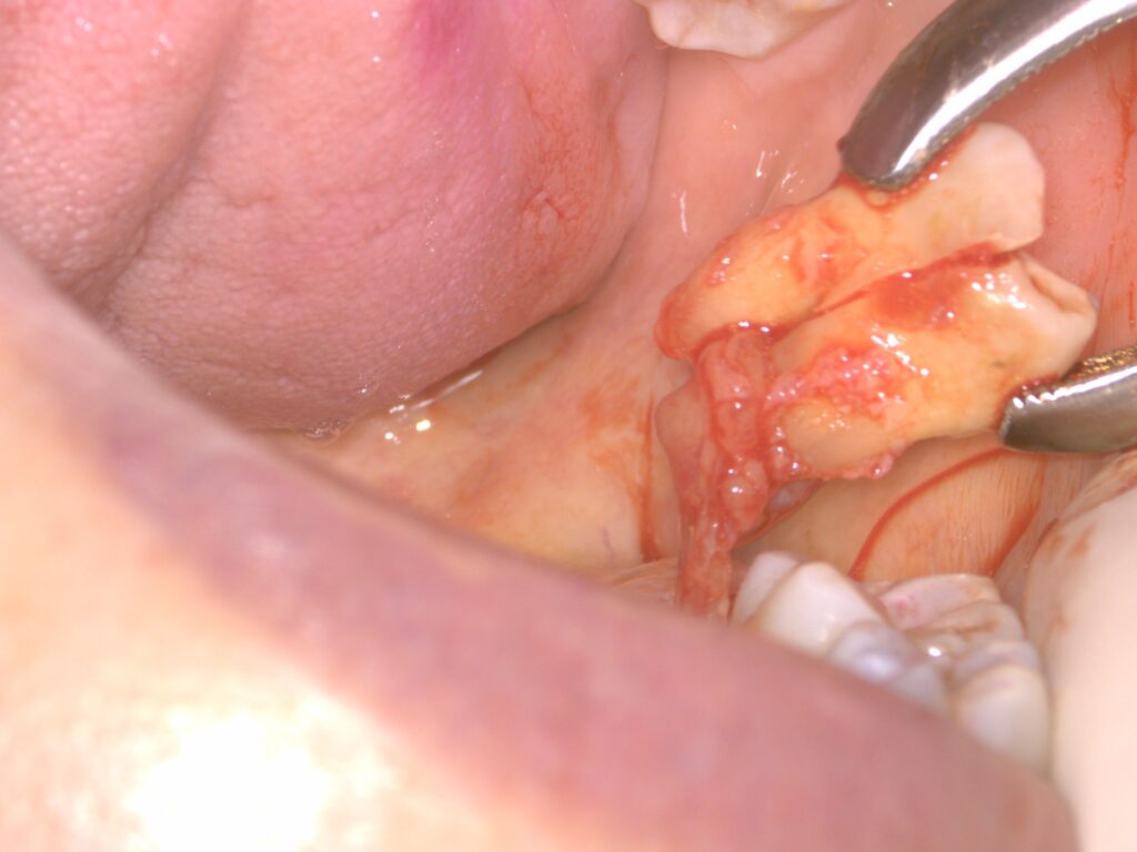
Please note: the following section contains photographs taken during surgery and photographs of the removed tooth.
Tooth-preserving treatment through intentional replantation
In this case, intentional replantation involved removing the tooth, treating the fracture and root outside the mouth as needed, and returning the tooth to its original position.
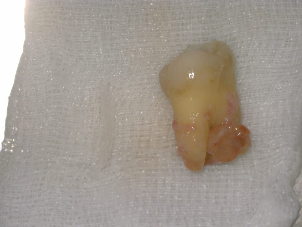
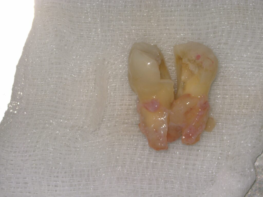
View more treatment photographs and follow-up for the upper right second molarHide additional treatment photographs and follow-up for the upper right second molarClick or tap to expand or collapse
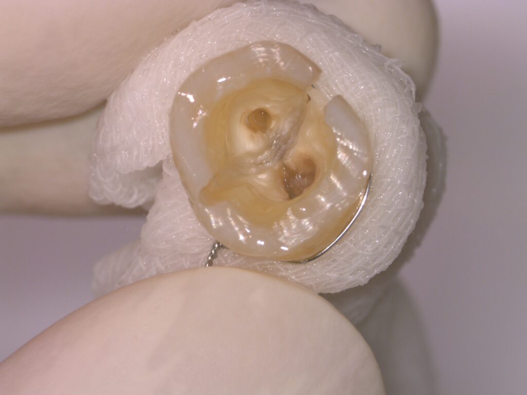
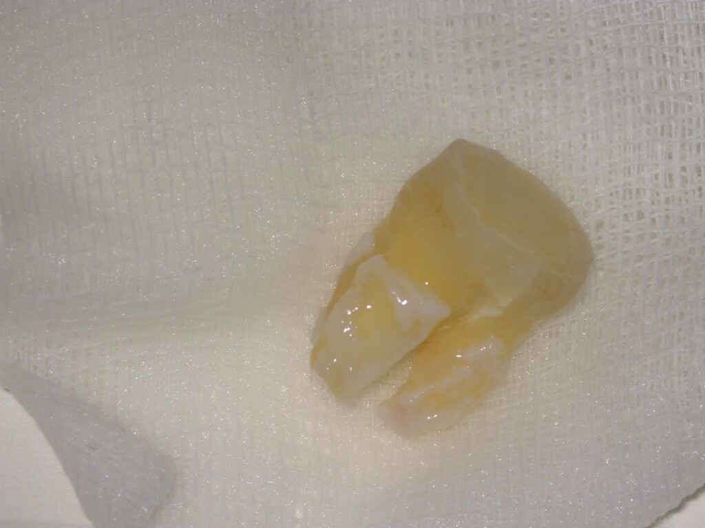
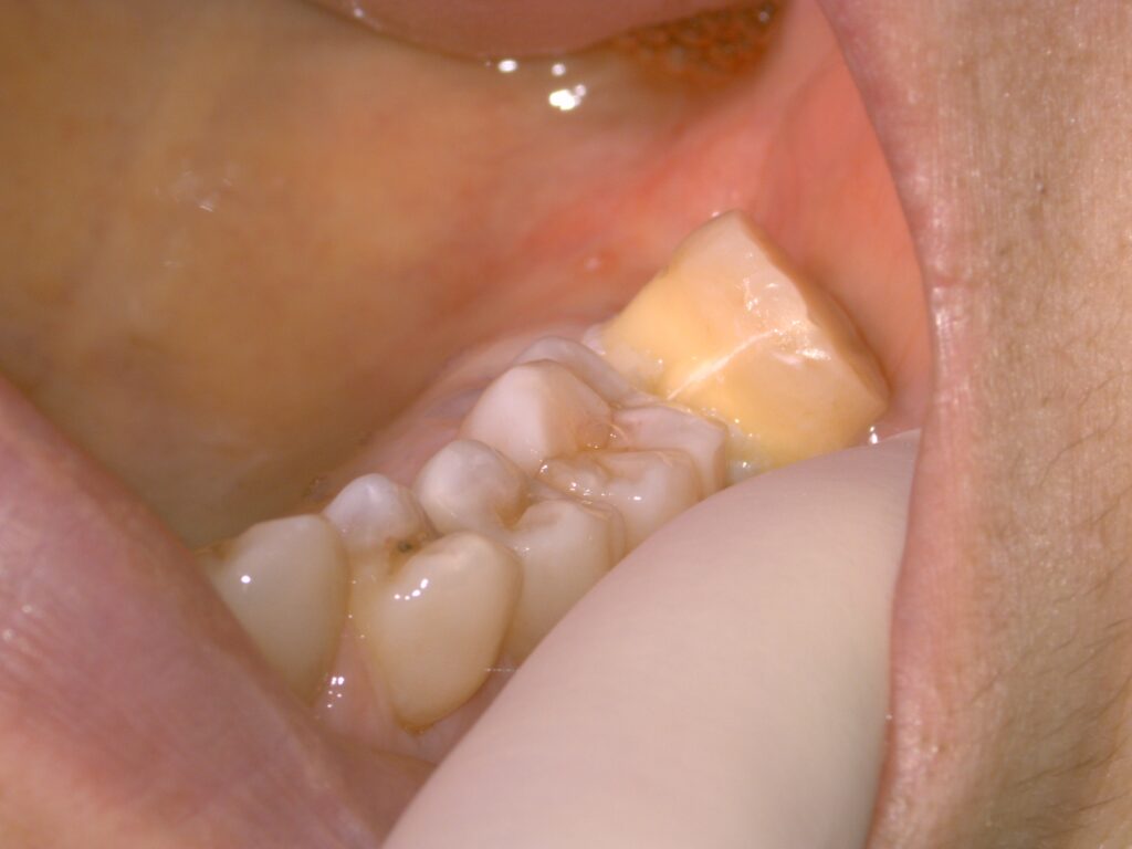
The fracture was difficult to see when the fractured surfaces of the removed tooth were together. Once separated, a fracture extending from the crown to the root was visible. Granulation tissue associated with inflammation was also observed around the root.
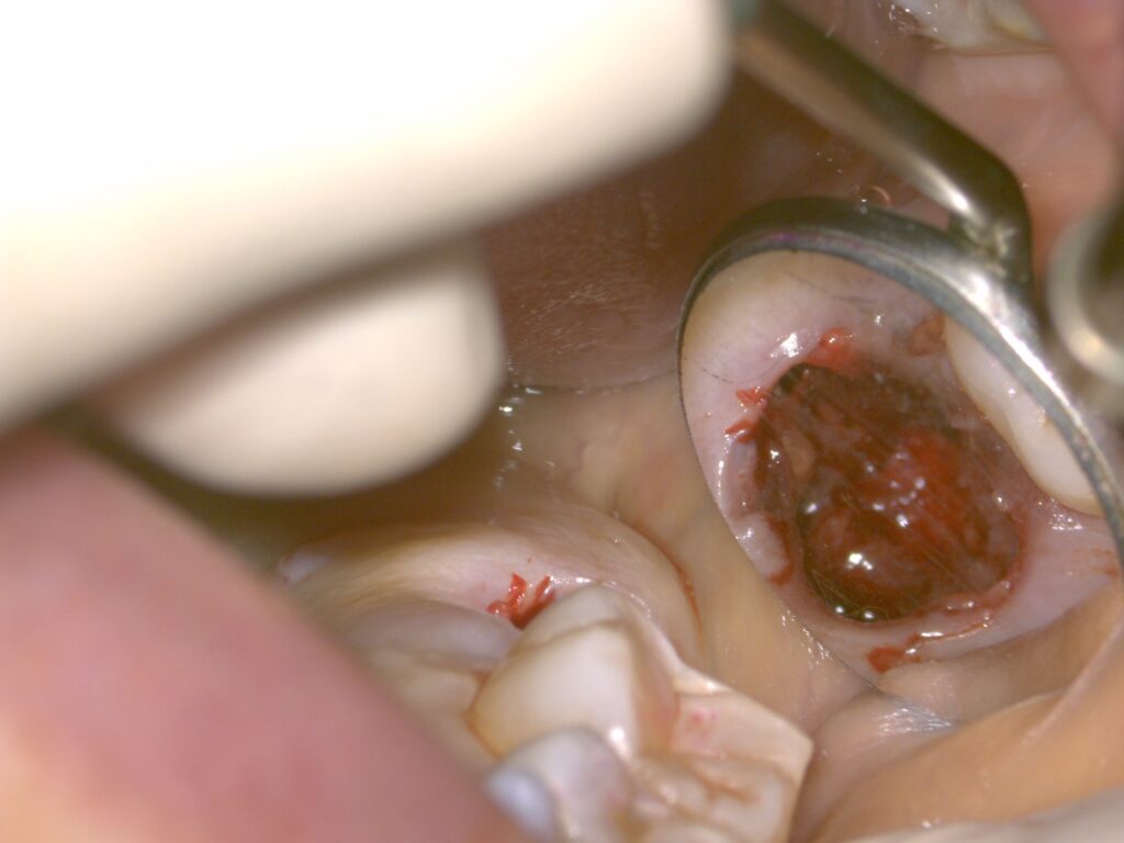
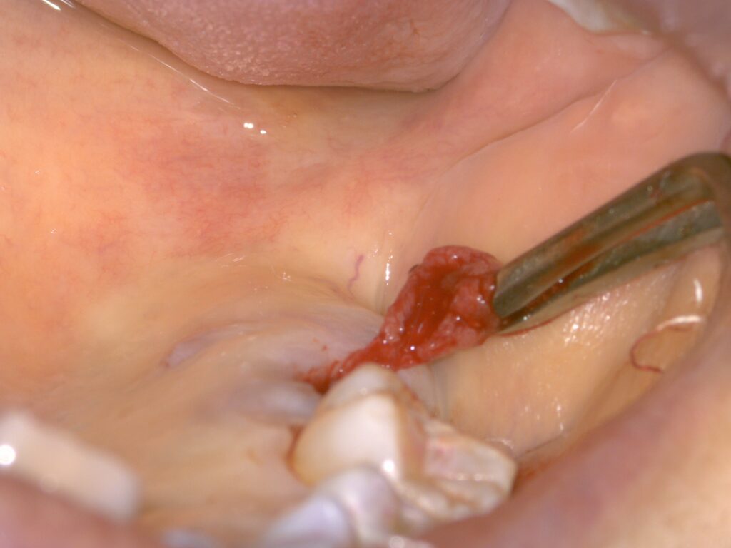
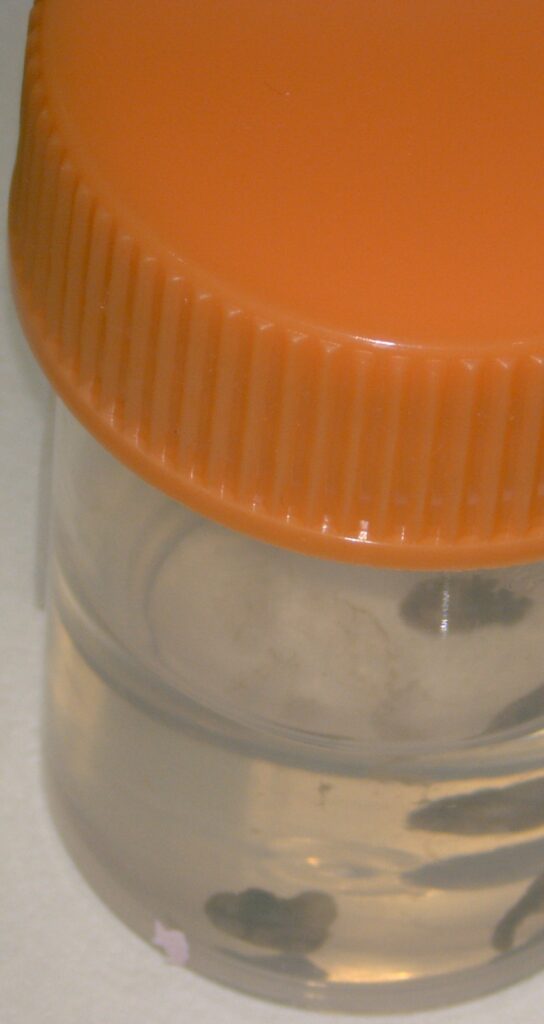
The surrounding tissues were treated under microscopic observation, and procedures including adhesive repair of the fracture were performed outside the mouth. The repaired tooth was then returned to its original position.
Splinting and follow-up after replantation
After the tooth was returned, it was splinted, and the tooth and surrounding tissues were monitored.
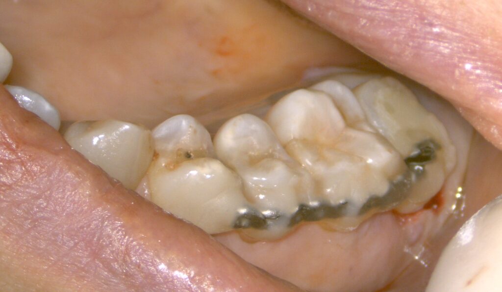
The splint was removed approximately 3 months after surgery. At that point, improvement in the gum condition and tooth mobility was observed. Improvement in the odontogenic maxillary sinusitis was also observed after treatment.
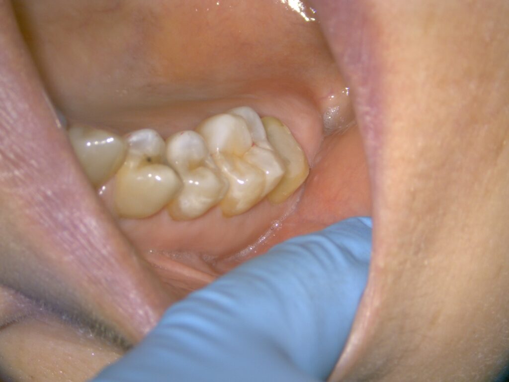
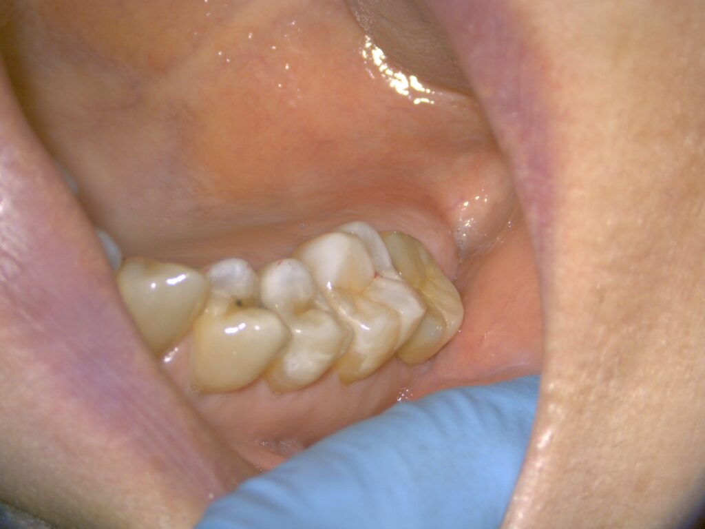
The time needed for inflammation to improve and for the tissues supporting the tooth to recover varies between individuals. Rather than ending treatment once the inflammation improved, restoration proceeded while the condition of the tooth continued to be assessed.
Placement of a zirconia crown
After assessing the postoperative condition, an impression was taken and a zirconia crown was made and fitted. The photograph shows the crown on a model before placement.
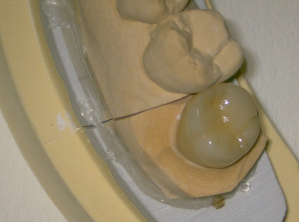
Whether a replanted tooth can be retained over the long term also depends on its subsequent course. Continued checks of the teeth, gums, and bite are important after treatment.
Treatment overview
Treatment site: upper right second molar (upper right 7)
Diagnosis: root fracture and odontogenic maxillary sinusitis
Main treatments: intentional replantation, adhesive repair of the fracture, treatment of infection, splinting after replantation, and restoration with a zirconia crown
Treatment duration: approximately 4 months
Number of visits: around 8
Cost: JPY 640,000 (tax included; private treatment not covered by health insurance)
Post-treatment course: improvement in odontogenic maxillary sinusitis was observed, and a zirconia crown was fitted
The cost, duration, and number of visits shown are specific to this case. They vary depending on the procedures required and the course of treatment.
Main risks and considerationsPress + to expand the explanationPress − to collapse the explanation
Intentional replantation is not suitable for every fractured tooth. The fracture or damage may extend during removal, making replantation impossible.
In addition to treatment-related pain, swelling, bleeding, and infection, possible complications after replantation include further fracture, reinfection, root resorption, fusion of the tooth to the bone, and poor healing of surrounding tissues. Crowns may also chip, break, or become detached.
If symptoms fail to improve or recur, additional treatment or extraction may be necessary. If inflammation in the maxillary sinus persists, care from an ear, nose, and throat specialist may also be required.
This case describes one patient’s treatment course and does not guarantee the same result or long-term tooth retention. During consultation, we explain the possibilities and limitations of preserving the tooth, the burden of treatment, and other options, including extraction.
The text and photographs for this case are published with the patient’s consent.
Case 2 | Root fracture in an upper right first premolar: adhesive repair and intentional replantation
A seemingly healthy tooth split in two: a case of repairing a root fracture discovered after gum swelling and pain
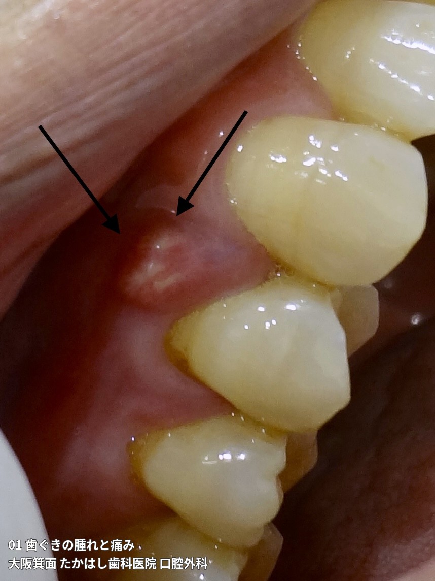
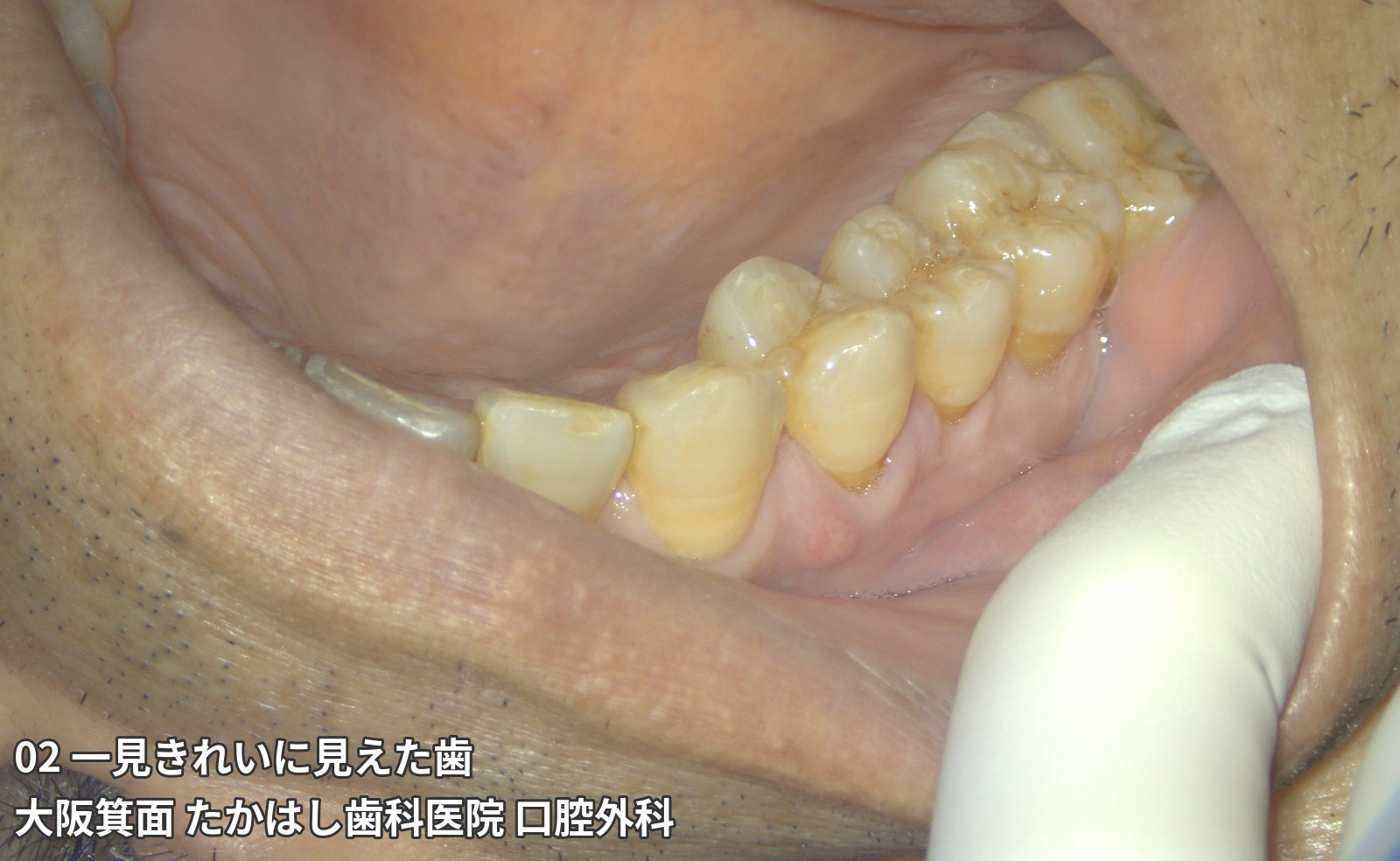
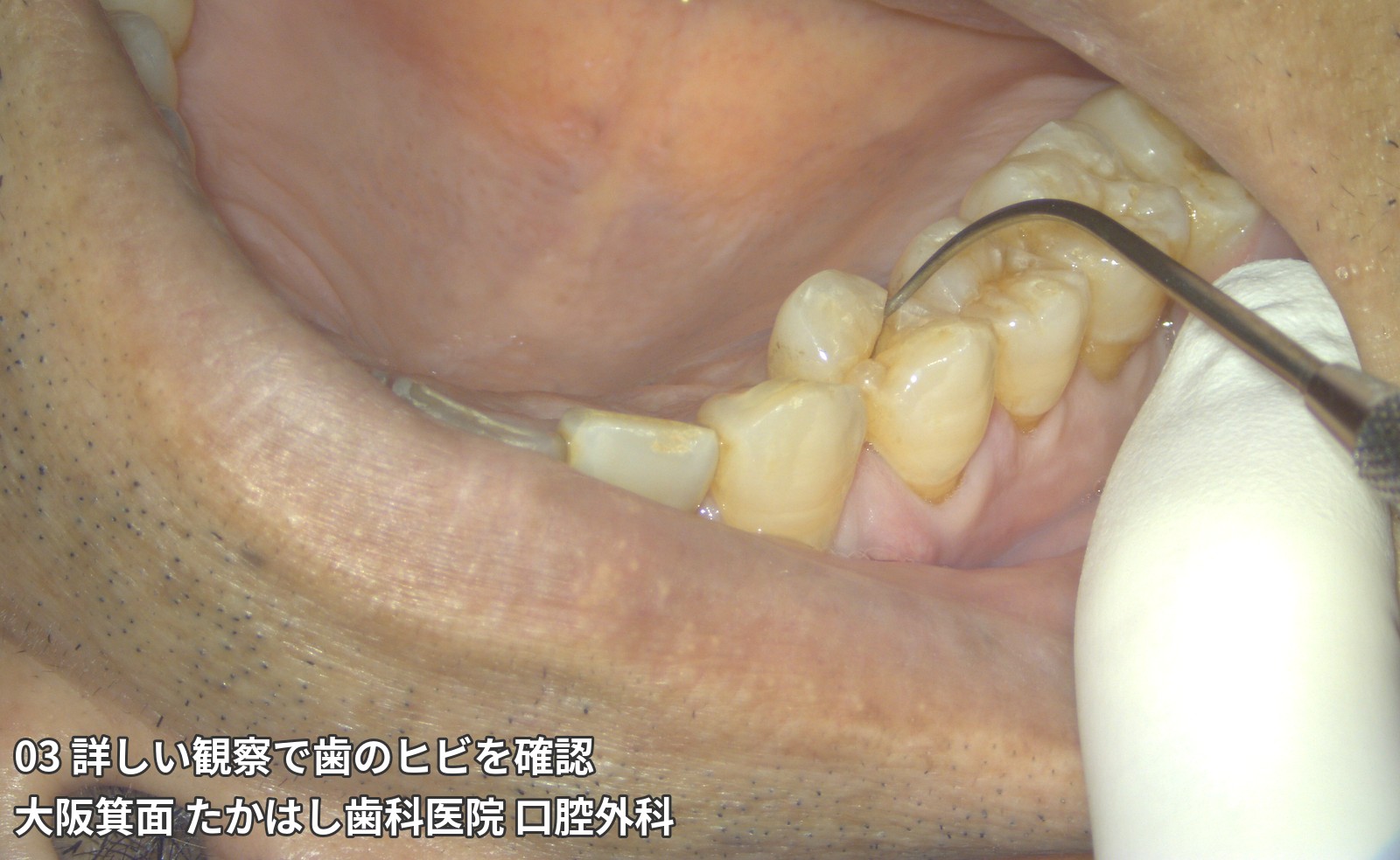
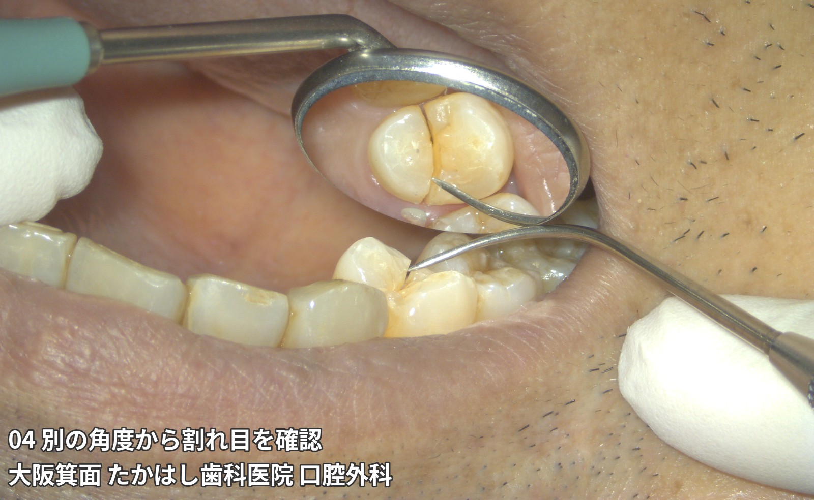
The patient presented with swollen gums and tooth pain. At first glance, the upper right tooth had no large cavity, but close examination revealed a fracture line (a crack or split). Intentional replantation was performed: the tooth was temporarily removed from the mouth, repaired with adhesive, and returned to its original position. A zirconia crown (a white dental crown) was subsequently fitted.
The affected tooth was the upper right 4 (upper right first premolar). Twelve photographs show the course from before treatment through replantation and zirconia crown placement. The following section includes treatment and intraoral photographs.
Treatment overview for this case:Adhesive repair, intentional replantation, and restoration with a zirconia crown. Private treatment not covered by health insurance, with a total cost of JPY 640,000 (tax included), 9 visits, and approximately 3 months until crown placement.
Main risks:Risks include pain and swelling after the procedure, infection, further fracture, root resorption, and fusion of the tooth to the bone. Depending on the course, it may not be possible to retain the tooth, and extraction may be necessary. The photographs show an individual case and do not guarantee the same result or long-term tooth retention.
01 Gum swelling and pain
02 A tooth that looked healthy at first glance
03 A crack identified on close examination
04 Examining the split from another angle
05 Removing the tooth from the mouth for repair
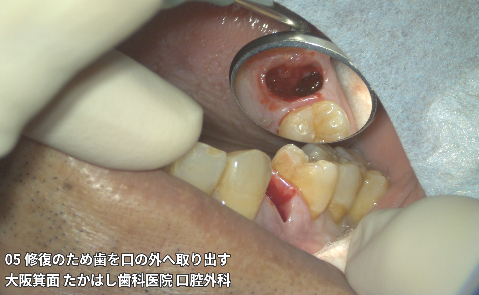
06 Examining the tooth outside the mouth
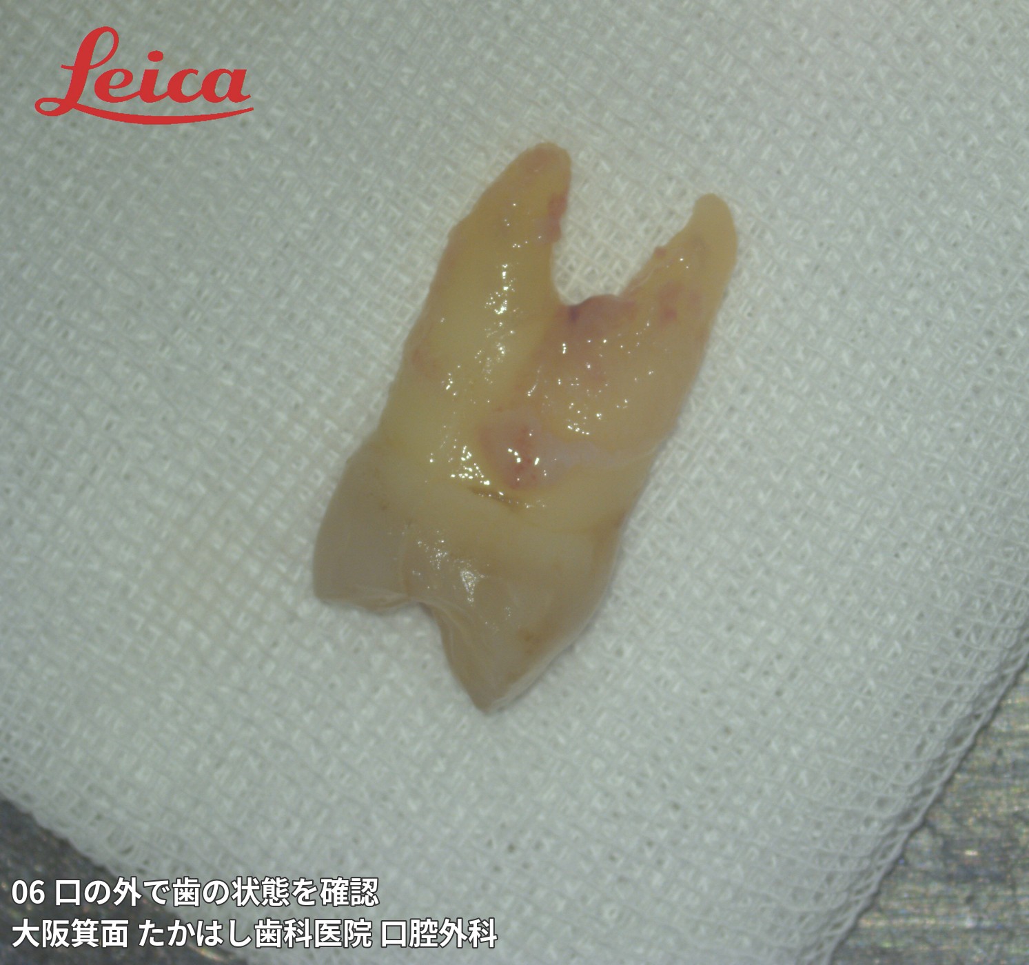
07 A tooth completely split in two
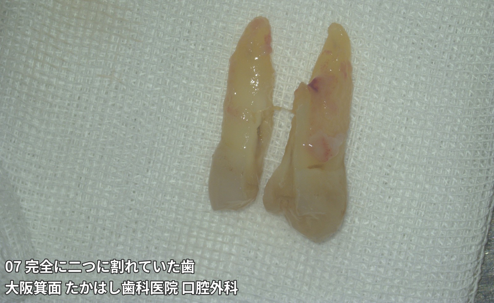
View more treatment photographs and follow-up for the upper right first premolarHide additional treatment photographs and follow-up for the upper right first premolarClick or tap to expand or collapse
08 Returning the repaired tooth to its original position
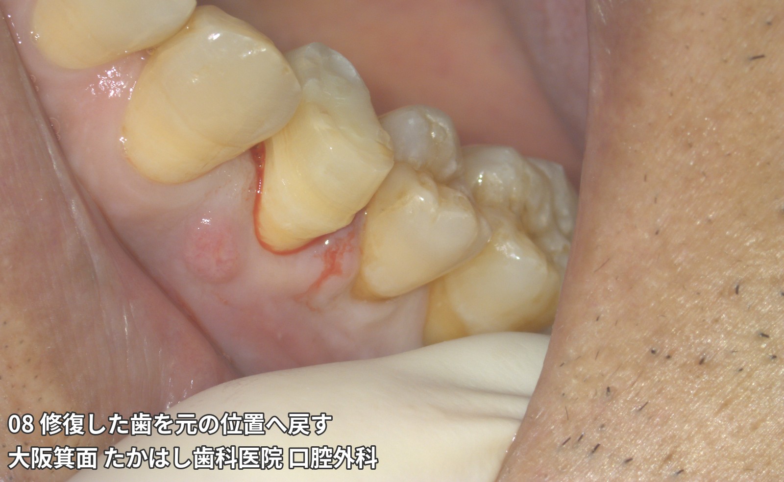
09 Splinting the replanted tooth
10 Removing the splint and preparing for a crown
11 Shaping the tooth and taking an impression
12 Placement of a zirconia crown
Whether a tooth can be saved is determined by examining the fracture pattern, infection, remaining tooth structure, surrounding bone, and other factors. Regular follow-up remains important after treatment.
When you are told, ‘Your tooth is fractured’ or ‘It needs to be extracted.’
Together, we explore the possibility of saving your tooth and the limits of treatment.
At Takahashi Dental Clinic, we consider tooth-preserving treatments such as adhesive repair and intentional replantation for root fractures and deep cracks in teeth. Our focus is not only on retaining a tooth, but also on how it can continue to function after treatment.
Not every tooth can be saved. Based on our examination, we explain what may be achievable, what may be difficult, and other options, including extraction.
Please note: the tooth-preserving treatments described on this site are private treatments not covered by health insurance. Intentional replantation involves removing a tooth, treating it, and returning it to its original position on the same day. The aim is to use your own tooth again.
What to consider before deciding on extraction
- You have been told that your tooth root is fractured and want to know whether it can be saved
- A previously treated tooth repeatedly develops swelling or pain when biting
- You want to discuss whether adhesive repair or intentional replantation is suitable for your tooth
- You want to compare the cost and burden of tooth-preserving treatment with treatment after extraction
We assess your current condition while listening to your wish to keep your own tooth if possible. Swelling and pain can have causes other than a root fracture, so symptoms alone cannot determine the treatment.
For those advised to have a tooth extracted at another clinic
What is a root fracture? A crack or split in the root of a tooth
A root fracture is a crack or split in the root that supports a tooth. The treatment needed varies depending on the location and extent of the fracture, infection, and the condition of the surrounding bone and gums.
A root fracture may require extraction. In some circumstances, however, tooth preservation may be worth considering. We carefully assess whether adhesive repair, intentional replantation, or other approaches are suitable. These treatments are neither appropriate for every root fracture nor a guarantee of long-term tooth retention.
Assessing the possibility of saving the tooth
During examination, we assess the fracture, periodontal tissues, infection, remaining tooth structure, bite, and other factors. CT scans and a dental microscope are used when needed to help determine a treatment plan.
Imaging alone cannot always confirm every fracture. Further examination or direct observation during treatment may change the initial outlook or treatment plan.
Treatments considered to preserve a tooth
Adhesive repair: aiming to seal and reinforce the fracture
After cleaning and preparing the fracture, adhesive materials are used to seal or reinforce it. We assess whether the fracture can be reached for treatment, whether infection can be controlled, and whether the load on the tooth after treatment can be adjusted.
Adhesive repair does not return the tooth to the same condition as an originally healthy tooth. Further fracture or recurrent infection may occur.
Intentional replantation: removing a tooth, treating it, and returning it on the same day
The tooth is removed, the root and fracture are treated outside the mouth as needed, and the tooth is returned to its original position on the same day. This approach may be considered when treatment is difficult to perform entirely inside the mouth.
Replantation may not be possible, for example if the fracture extends during removal. Infection, root resorption, fusion of the tooth to the bone, and other complications after replantation may also prevent a stable course.
Learn about intentional replantation
Related treatments according to the condition
Treatment may be combined with surgical endodontic treatment for a lesion at the root tip, removal of part of the roots in a multirooted tooth, or bite adjustment. Depending on the condition of the surrounding bone, bone regeneration treatment may also be considered.
Rather than performing all procedures routinely, we explain the need, expected benefit, and additional burden of each.
View treatment and cost details
When preservation is difficult, we explain the reasons and options
Depending on the extent of the fracture, severity of infection, and condition of the tissues supporting the tooth, it may be difficult to retain the tooth even after repair. The tooth’s anatomy, general health, or other factors may also prevent a planned surgical procedure.
After attempting tooth-preserving treatment, the plan may need to change to extraction during treatment or follow-up. Continuing to insist on preserving a tooth is not always appropriate for the surrounding tissues.
If extraction is necessary, we explain why and discuss options such as implants, bridges, and dentures. We compare costs, treatment duration, effects on adjacent teeth, and maintenance to help you decide on a plan you feel comfortable with.
From consultation to follow-up care
1. We discuss your history and wishes
Please tell us about your symptoms, previous treatment, why extraction was recommended, your wishes, and any circumstances affecting your ability to attend appointments. If you have a referral letter or previous imaging, please let us know when booking.
2. We examine the tooth and assess the prospects for preservation
Based on an oral examination and any necessary tests, we assess the suitability and limitations of treatment. We distinguish what can be determined at the time of examination from what remains uncertain.
3. We explain the treatment plan, costs, and risks
We explain the proposed procedures, alternatives, expected visits, costs, and circumstances that may lead to additional charges. We address your questions and proceed once you understand and agree to the plan.
4. We carry out treatment and assess recovery and function
As we monitor the condition after treatment, we adjust the bite, crown, or other aspects as needed. The plan may be revised depending on the treatment and subsequent course.
5. We check for changes at regular follow-up visits
After treatment, we continue to assess the teeth, gums, bite, and changes on imaging. We also provide guidance on cleaning and, when needed, use of a night guard.
The course is influenced by multiple factors, including the tooth’s condition, treatment, healing response, and biting forces. Recurrence or tooth loss remains possible even with continued home care and regular dental visits.
Private treatment: costs, duration, and risks
The tooth-preserving treatments described on this site are private treatments not covered by health insurance. The procedures needed depend on the tooth’s condition. We explain the treatment and costs after examination.
- Intraoral adhesive repair: Base fee of JPY 550,000, including tax and the crown. Additional charges may apply depending on the condition of the tooth. Treatment pathway: Initial consultation, a private-treatment agreement at the second appointment, treatment at the third appointment, impressions around the fourth or fifth appointment, and final crown fitting. The overall course is approximately 1–3 months.
- Intentional replantation: Approximately JPY 640,000 per tooth, including tax and the crown. Additional charges may apply depending on the condition of the tooth. Treatment pathway: Initial consultation, a private-treatment agreement at the second appointment, surgery, and postoperative checks at 1, 2, 4 and 8 weeks. Impressions are taken at 12 weeks, with the final restoration fitted at a later appointment.
- Maintenance: Every 1–6 months (risk-based maintenance)
Main risks and side effects
- Procedures may cause pain, swelling, bleeding, infection, or other complications.
- Further fracture or reinfection may occur after adhesive repair.
- With intentional replantation, damage during removal or other factors may make replantation impossible. Root resorption or fusion of the tooth to the bone may occur after replantation.
- Depending on the condition and treatment, there may be individual risks, such as effects on nerves or surrounding tissues.
- Symptoms may fail to improve or may recur after treatment, and additional treatment or extraction may be necessary.
Suitability, treatment duration, and the subsequent course vary between individuals. We explain the risks specific to the planned procedure and the available options, including no treatment.
Frequently asked questions
Can a tooth diagnosed with a root fracture always be saved?
Not always. We examine the fracture and surrounding tissues to determine whether preservation can be considered. If the prospects for saving the tooth are poor, we explain why.
When is the removed tooth returned during intentional replantation?
The tooth is removed, treated as needed, and returned to its original position on the same day. Unlike treatment that replaces a tooth with an artificial tooth after a conventional extraction, the aim is to use your own tooth again. However, damage during removal or other factors may make replantation impossible.
Which is better: preserving the tooth or an implant?
There is no single answer for everyone. We compare the prospects and burden of preserving the tooth with the advantages and disadvantages of treatment after extraction. While respecting your wish to keep your own tooth, we explain options suited to the condition of your whole mouth.
Can I consult you if I live far away?
Yes. However, visits to the clinic are necessary for examination and follow-up after treatment. The number and spacing of visits vary by case, so please tell us where you live and any circumstances affecting travel when booking.
View more frequently asked questions
A consultation to help you make an informed choice about a valued tooth
We aim to respect both your wish to keep your tooth and the clinical outlook. Let us first assess its current condition and clarify whether tooth-preserving treatment may be considered, along with the burden and risks involved.
Takahashi Dental Clinic
1F, 4-16-33 Boshima, Minoh, Osaka, Japan
Phone: 072-725-1182
Related information
- Root fracture treatments and costs
- About adhesive repair
- About intentional replantation
- Treatment cases Please note: these are individual cases and do not guarantee similar results.
- For those deciding between extraction and preservation, including conditions other than root fractures (main clinic website)
- Takahashi Dental Clinic main website