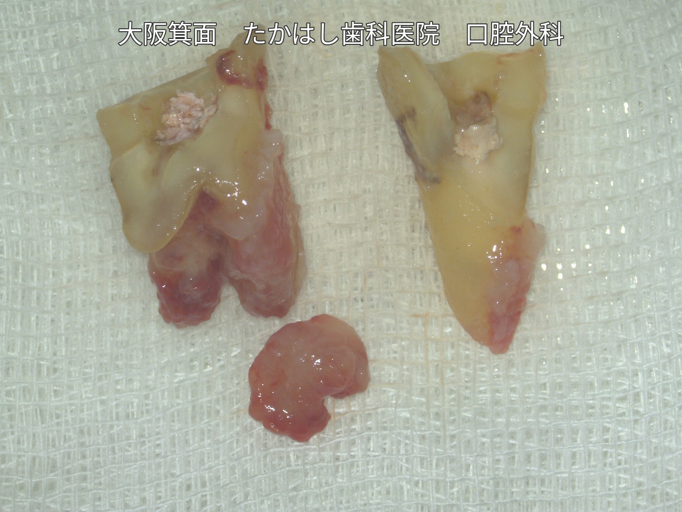
Adhesive repair and replantation of an upper right molar with a vertical fracture extending to the root tip
This case involved an upper right first molar with a vertical fracture extending from the crown to the root tip. Photographs show extraoral adhesive repair, root canal treatment, root-end resection, replantation and splinting, through to zirconia crown placement.
Case video
Treatment, cost, duration and number of visits for this case
Treatment: Extraoral adhesive repair, root canal treatment and root-end resection, replantation and wire splinting, and final restoration with a zirconia crown.
Total cost: JPY 640,000 (tax included, including placement of the final zirconia crown)
Treatment duration: Approximately 3 months
Number of visits: 9 visits
The visits consisted of the initial consultation, an explanation of the treatment, surgery, follow-up at 1, 2, 4 and 8 weeks after surgery, an impression at 12 weeks, and crown placement. Treatment duration and the number of visits vary by case.
Main risks and treatment limitations
Reinfection, root resorption, ankylosis (fusion of the tooth to bone), or poor healing after replantation may occur, and further treatment or extraction may be necessary. Outcomes vary from person to person; preservation of the tooth and its long-term prognosis are not guaranteed. This case record covers the period up to placement of the final crown.
Condition before treatment and examination of the vertical fracture
The sequence begins with a photograph of the fractured upper right first molar before treatment. After removal and examination, a vertical fracture extending from the crown to the root tip was found. The fractured tooth was opened to examine its interior.
Extraoral repair and replantation
Adhesive repair, root canal treatment and root-end resection were performed outside the mouth. The repaired tooth was returned to its original socket and secured with wire to prevent displacement.
From postoperative follow-up to the final restoration
At 3 months after surgery, progress was checked, the splinting wire was removed and an impression was taken. A zirconia crown was subsequently fitted. The final photograph shows the final restoration in place.
The case photographs and video are used with the patient’s consent to publication.