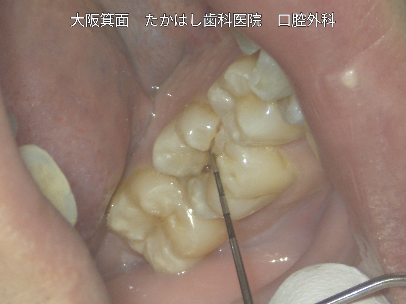
Adhesive repair without extraction of a lower right molar that fractured while eating and caused severe pain
The patient presented with severe pain after the lower right first molar fractured while eating. In this case, adhesive repair was performed inside the mouth rather than removing the tooth for extraoral repair. Photographs show the sequence from root canal treatment and glass-fiber core build-up through zirconia crown placement.
Case video
Treatment, cost and duration for this case
Treatment: Microscope-assisted intraoral adhesive repair, root canal treatment, glass-fiber core build-up and zirconia crown placement.
Cost: JPY 550,000 in total (tax included)
Treatment duration: Approximately 1 month
Number of visits: 3
The JPY 550,000 fee includes microscope-assisted intraoral adhesive repair, root canal treatment, glass-fiber core build-up and the restoration (zirconia crown). The cost, duration and number of visits refer to this individual case.
Main risks and treatment limitations
Reinfection, progression of the fracture or refracture may occur after treatment, and further treatment or extraction may be necessary. Outcomes vary depending on the location and extent of the fracture, the condition of the tooth and periodontal tissues, and other factors. This is one individual case and does not guarantee that the same treatment is suitable for every tooth, that the tooth can be preserved, or that long-term treatment outcomes will be achieved.
Condition at presentation and treatment plan
The tooth fractured while the patient was eating. Because it still contained vital pulp, it caused severe pain. The fracture was recent and contamination was considered limited, so intraoral repair without extraction was selected for this case.
Rubber dam placement and microscope-assisted treatment
A rubber sheet called a rubber dam was placed to prevent saliva from entering the tooth. The fractured area was carefully removed under microscopic observation. This is a time-consuming procedure requiring close examination of fine details.
Adhesive repair and root canal treatment
The fractured area was repaired with Super-Bond. After treatment of the pulp, the root canals were filled. The corresponding photograph in the video shows the tooth after root canal filling, with the pink filling material visible.
Glass-fiber core build-up and crown placement
Four or more glass-fiber posts were used to build up the core and reinforce the inside of the tooth. The core was then shaped, an impression was taken and a zirconia crown was fitted.
This video records the treatment sequence up to crown placement. It does not document long-term follow-up. The case photographs and video are used with the patient’s consent to publication.