Preservation of a Root-Fractured Tooth Requiring Differentiation from Gingival Cancer
A case combining adhesive repair and intentional replantation with periodontal regenerative therapy
This case involved marked gingival swelling and bleeding, requiring differentiation from gingival cancer. Following examination, we diagnosed inflammatory epulis associated with a vertical root fracture that had completely separated the tooth. We preserved the tooth by bonding the fractured parts outside the mouth and combining intentional replantation with periodontal regenerative therapy.
- History before presentation
- Imaging assessment of the root and surrounding bone
- Histopathological diagnosis of inflammatory epulis
- Removal of the separated root fragments and extraoral adhesive repair
- Curettage of the extraction socket and periodontal regenerative therapy
- Imaging at four months after surgery
- Crown restoration and postoperative condition
- Key considerations in this case
- Treatment cost and visits for this case
- Limitations and main risks of tooth-preserving treatment
History before presentation
The patient was a woman in her 40s. She presented with swelling around the upper right first premolar and pain even at rest. The crown had come off approximately six months earlier, and another clinic had recommended extraction. However, she wished to keep her own tooth. She visited our clinic after the gum in the same area gradually became more swollen.
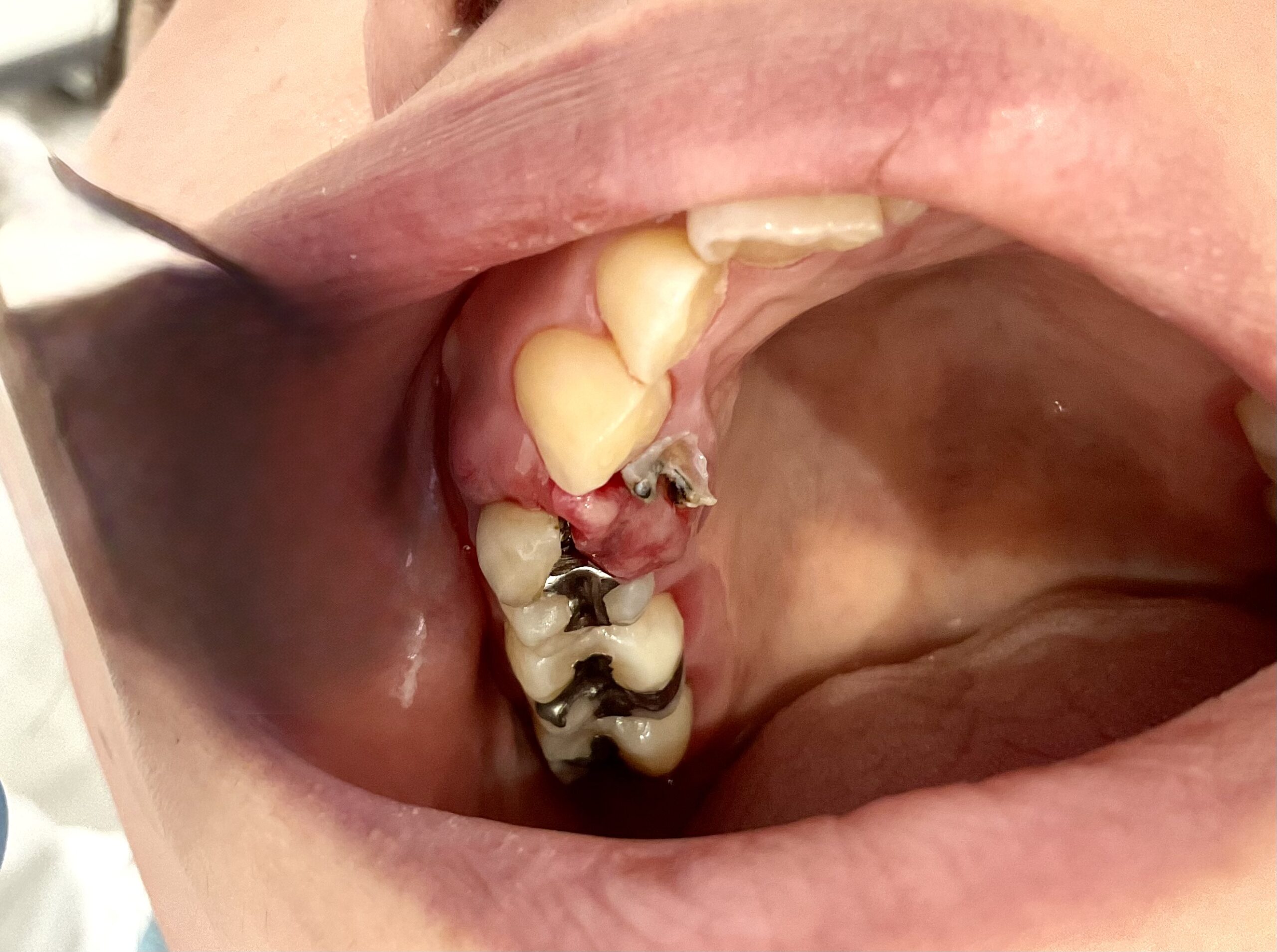
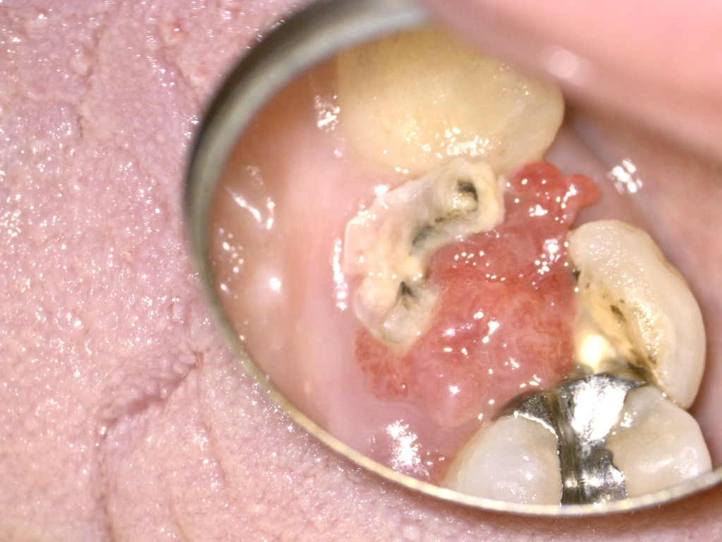
At the initial visit, there was a gingival mass with ulceration and bleeding. Rather than assuming that it was solely inflammation caused by the root fracture, we investigated the possibility of a malignant condition, including gingival cancer.
This page contains actual intraoral photographs, photographs taken during surgery, and photographs of specimens. The course of treatment is specific to this case; the same treatment and results do not apply to every patient.
Imaging assessment of the root and surrounding bone
We assessed the affected root and surrounding bone using panoramic radiography and dental cone-beam computed tomography (CBCT). We reviewed CBCT sections in multiple planes and evaluated the extent of the root fracture and bone defect together with the findings from the intraoral examination.
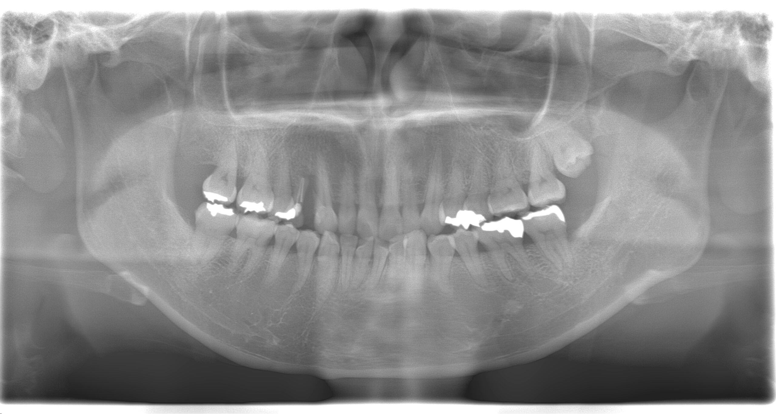
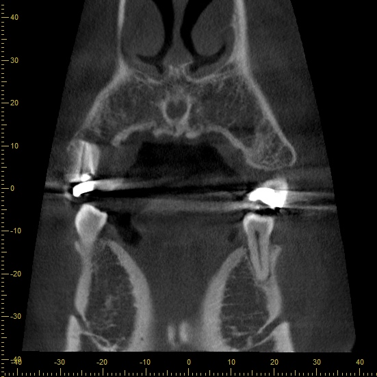
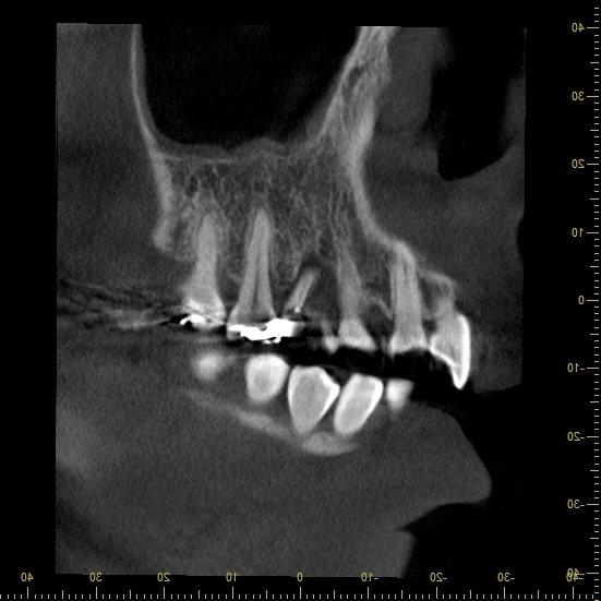
Imaging helped us assess whether the fractured tooth could be preserved. However, to establish the nature of the gingival mass, we also needed to take a tissue sample for histopathological examination.
Histopathological diagnosis of inflammatory epulis
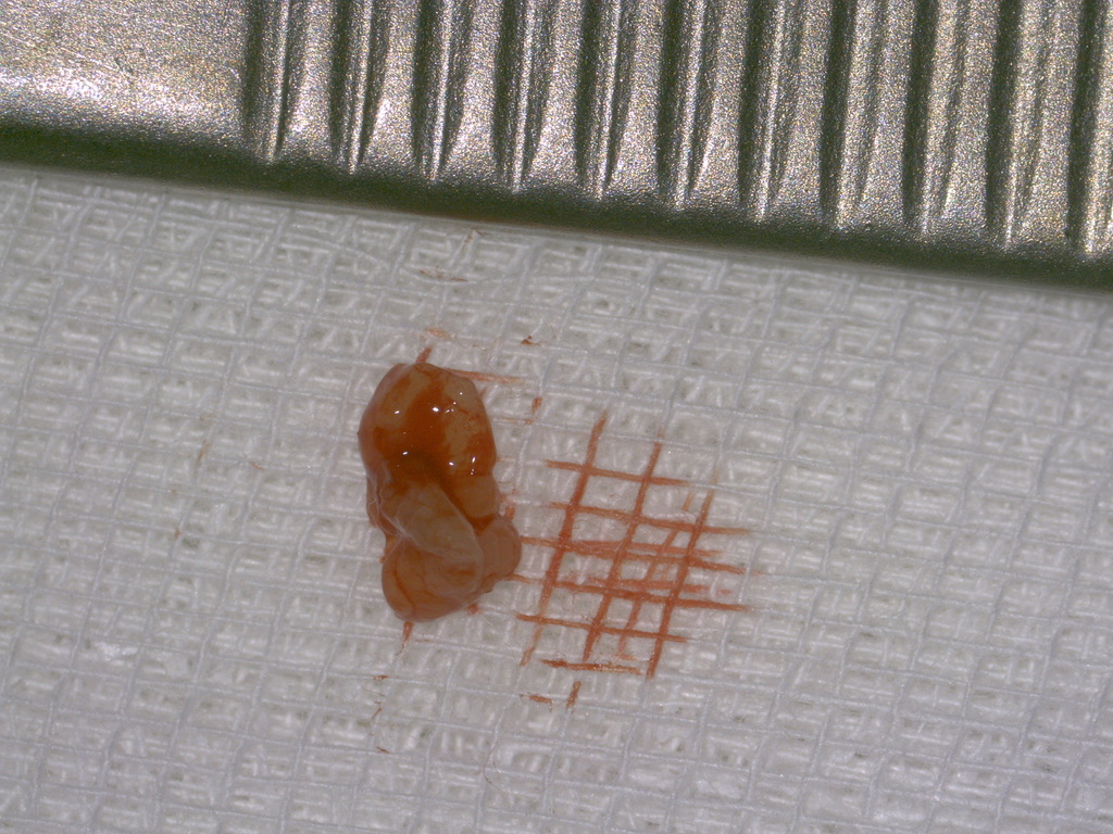
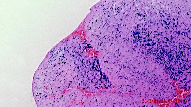
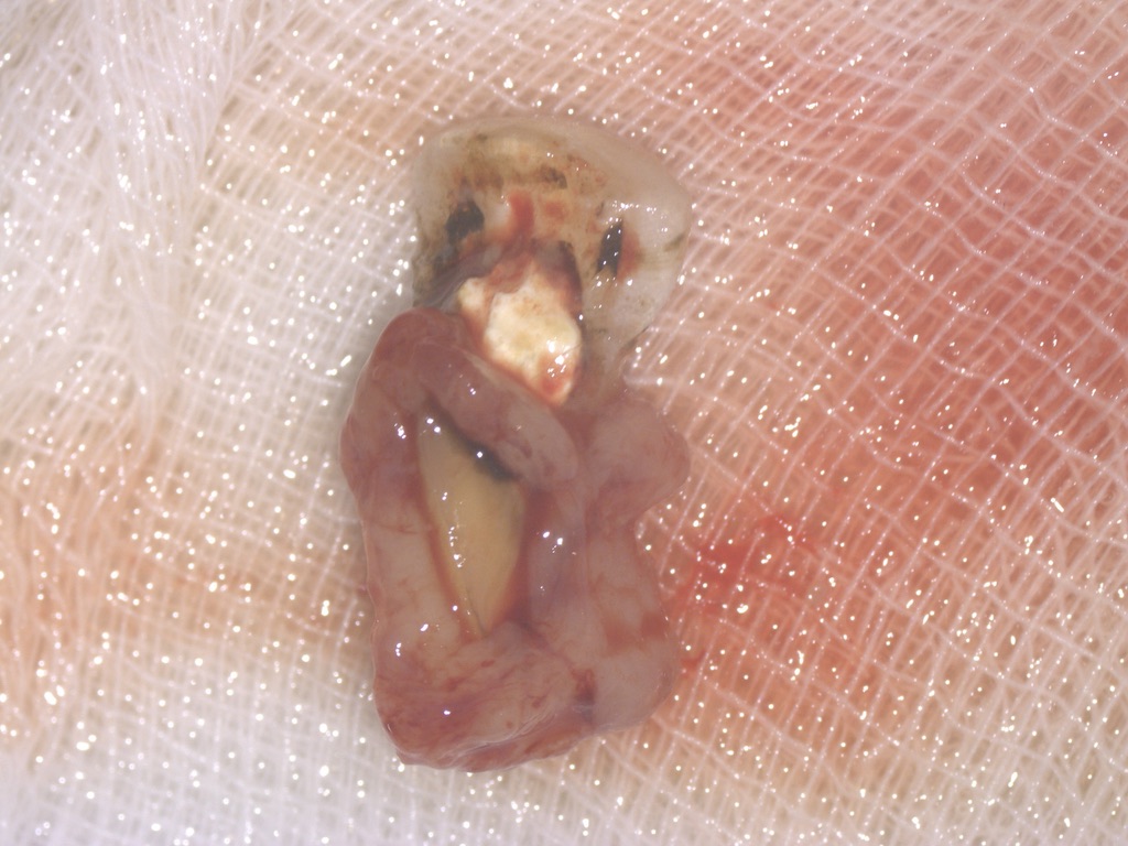
We performed an excisional biopsy of the gingival lesion and submitted it for histopathological examination. The diagnosis was inflammatory epulis. Epulis is a term for a localized swelling of the gum. In this case, chronic irritation from the completely separated root fragments was considered to have contributed to the reactive gingival lesion.
Removal of the separated root fragments and extraoral adhesive repair
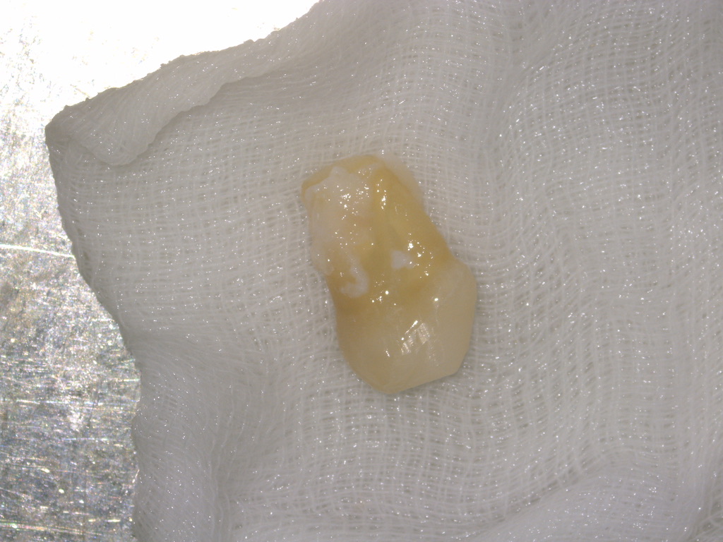
We selected intentional replantation after considering the diagnosis of the lesion, the condition of the tooth, and the patient’s wish to preserve it. Intentional replantation involves temporarily removing a tooth, carrying out the necessary treatment outside the mouth, and returning it to its original position. In this case, we removed the fractured tooth and the lesional tissue and bonded the fractured surfaces together.
Curettage of the extraction socket and periodontal regenerative therapy
The extraction socket is the space previously occupied by the tooth. We removed the lesional tissue from the socket, cleaned and curetted it, and used periodontal regenerative therapy to address the surrounding bone defect. In this case, we used bone graft material and trafermin with the aim of regenerating the periodontal tissues.
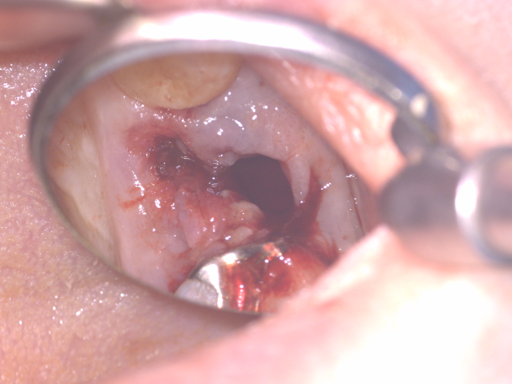
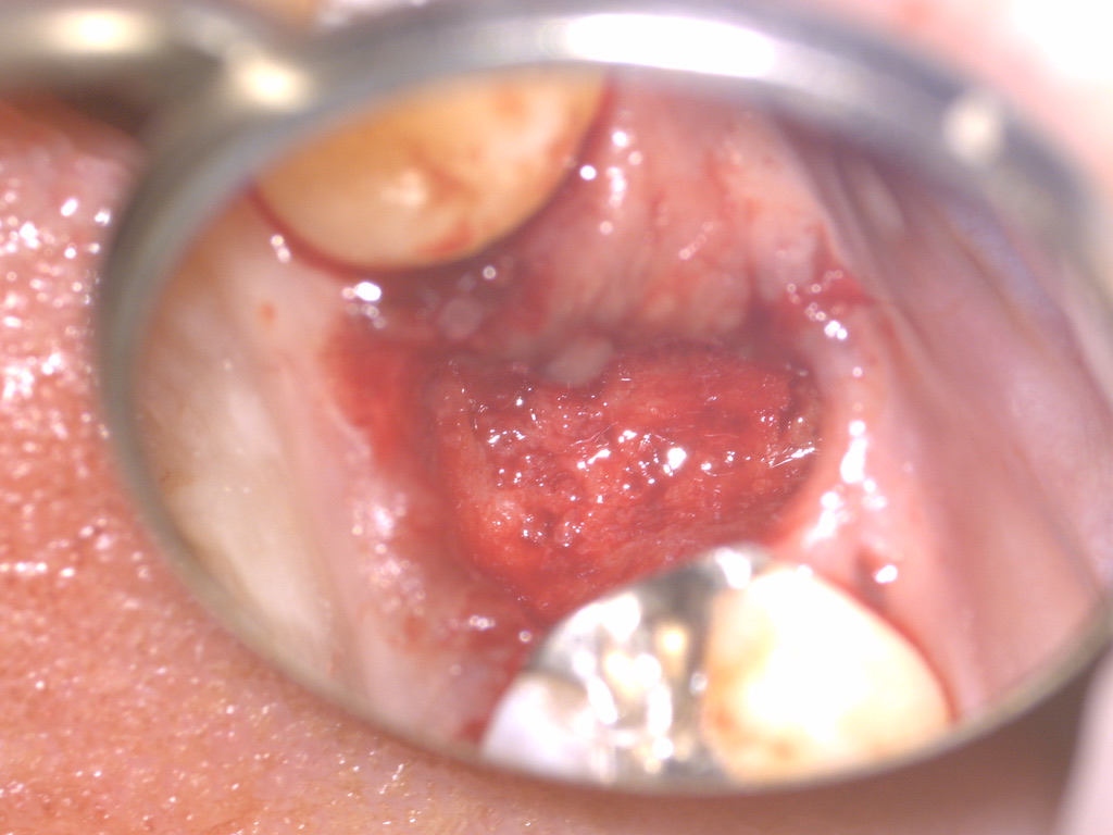
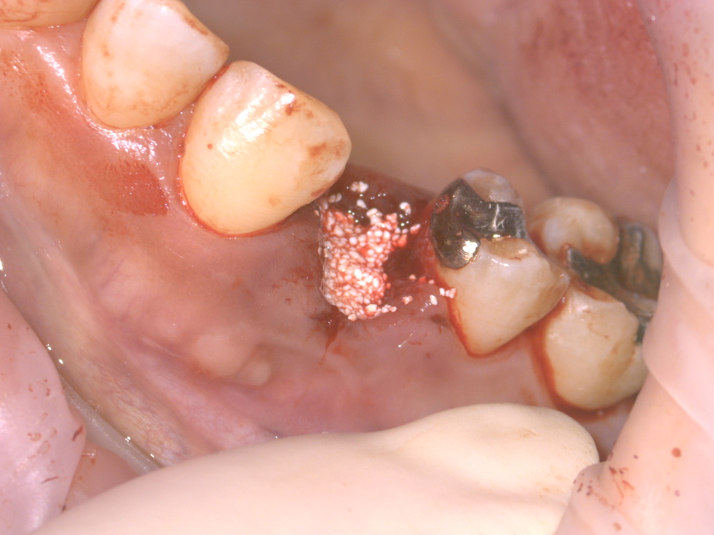
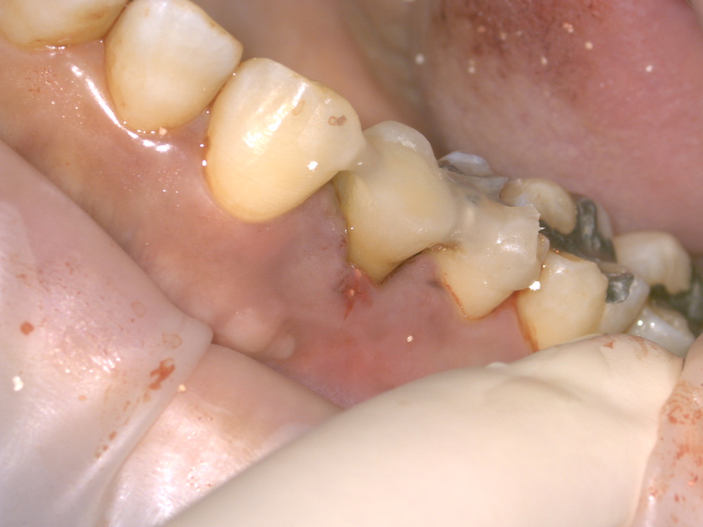
The adhesively repaired tooth was returned to its original position and stabilized on the day of the procedure. We then proceeded with bite management and prosthetic treatment while monitoring the tooth and surrounding tissues. Although replantation was completed that day, the entire course of treatment was not completed in a single day.
Imaging at four months after surgery
We assessed progress using panoramic radiography and CBCT four months after surgery. We evaluated the replanted tooth and surrounding tissues and continued to preserve the tooth.
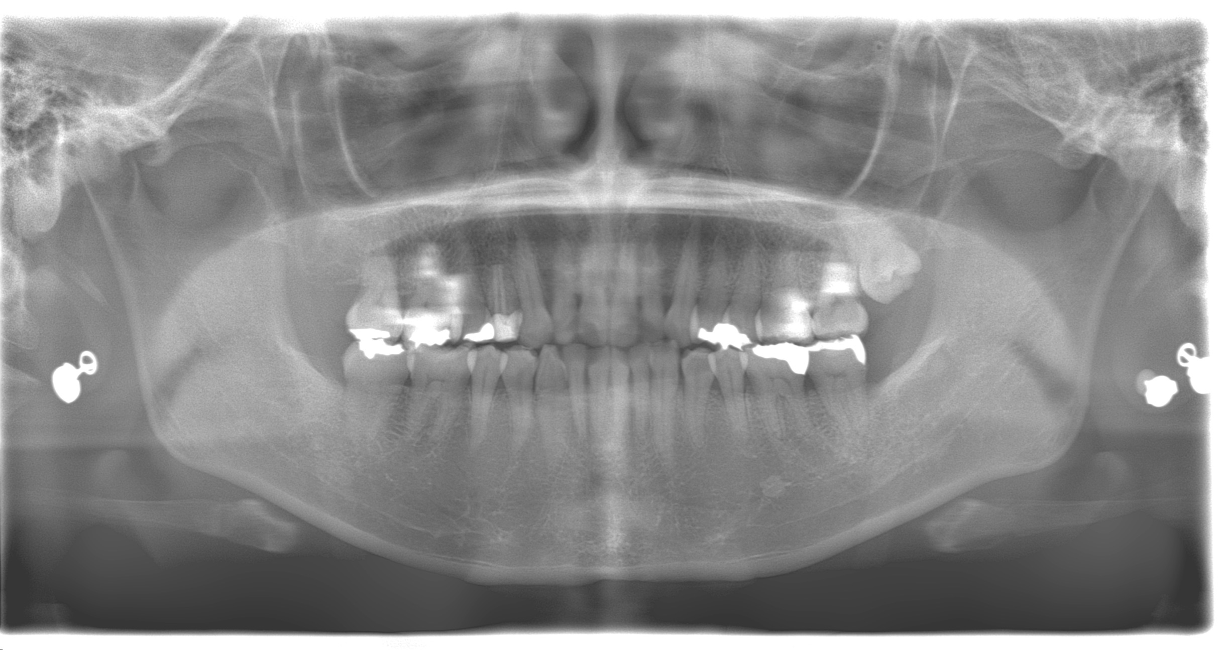
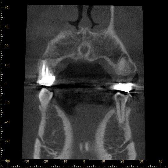
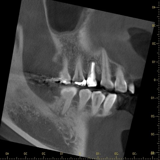
The planes and angles of the preoperative and postoperative CT images differ. Simply superimposing the images shown here cannot measure the amount of bone recovery. The condition after treatment is assessed using both clinical examination and imaging.
Crown restoration and postoperative condition
After assessing progress following replantation, we proceeded with crown restoration. The images below show the record made at the impression stage, the tooth before crown placement, and intraoral views after placement.
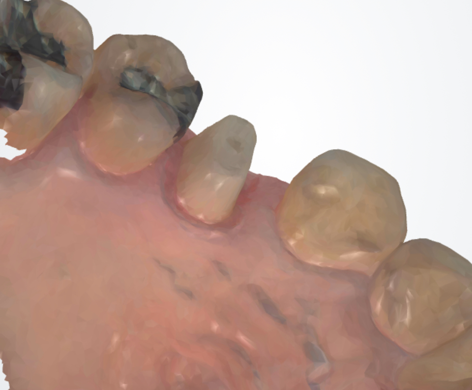
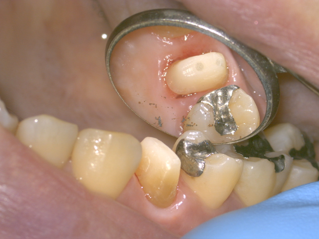
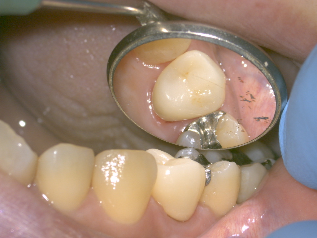
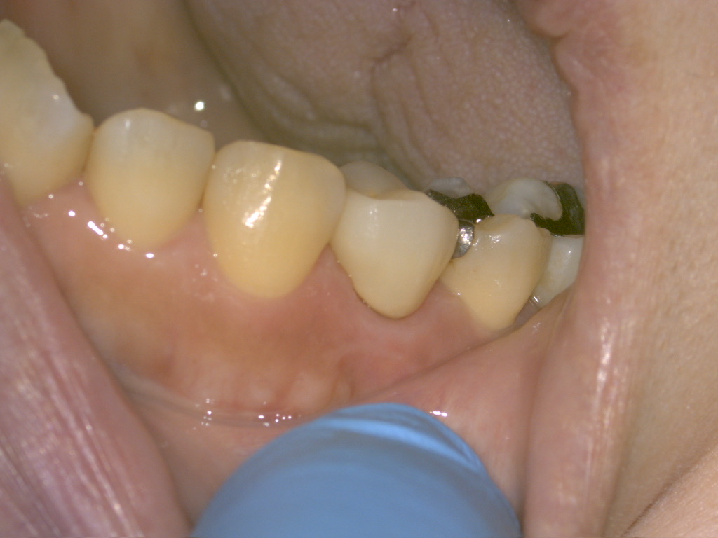
In addition to treating the gingival lesion, we carried out repair of the fractured tooth, replantation, recovery of the surrounding tissues, and restoration of function with a crown as a continuous course of treatment. Ongoing assessment of the tooth, periodontal tissues, and bite remains important after tooth preservation.
Key considerations in this case
This case required both an appropriate diagnosis of a lesion suspicious for gingival cancer and an assessment of whether the completely separated fractured tooth could be preserved. After establishing the nature of the lesion, we combined oral surgical diagnosis and treatment with adhesive repair of the tooth, intentional replantation, and periodontal regenerative therapy.
The key to treatment in this case was to address the underlying root fracture as well as removing the gingival mass. This does not mean that every tooth with a root fracture can be preserved. Suitability is assessed for each individual tooth.
Treatment cost and visits for this case
| Treatment cost | JPY 640,000 |
|---|---|
| Treatment duration | Approximately 4 months |
| Number of visits | 10 visits |
The cost, duration, and number of visits above are based on the treatment provided in this case. These vary according to examination findings and the procedures required and may not match the estimates in our general treatment information.
Limitations and main risks of tooth-preserving treatment
Whether a tooth can be preserved depends on the extent of the fracture, the infection, the remaining tooth structure, and the condition of the supporting bone and periodontal tissues. Even after tooth-preserving treatment, further treatment or extraction may become necessary depending on the course of recovery.
In addition to pain, swelling, bleeding, and infection associated with treatment, risks include damage during removal of the tooth, refracture, reinfection, root resorption, ankylosis (fusion of the tooth to the bone), and tooth mobility. After surgery, ongoing care includes oral hygiene and bite management, regular examinations, and imaging when necessary.
If gingival swelling or bleeding persists, do not judge the cause by appearance alone. Please seek an examination at a dental clinic or oral surgery department.
This article presents the course of a single case. It does not guarantee suitability for treatment or treatment outcomes.