This case involved a lower left first molar with only the roots remaining. Intraoral repair, core build-up using glass-fiber posts and placement of a provisional crown were followed by final restoration with a zirconia crown. The lower left first molar is the sixth tooth from the front on the lower left.
This is a case record from Takahashi Dental Clinic and Oral Surgery in Minoh, Osaka. This page contains photographs taken inside the mouth during treatment.
- 01 | Before treatment: Only the roots of the lower left molar remained
- 02 | Exposing the tooth covered by gum tissue and treating decay
- 03 | Repair inside the root canals
- 04 | Core build-up using glass-fiber posts
- 05 | Placement of a provisional crown on the same day
- 06 | Fabrication of a zirconia crown
- 07 | Zirconia crown placement
- Treatment, cost, duration and number of visits for this case
- Main risks and treatment limitations
01 | Before treatment: Only the roots of the lower left molar remained
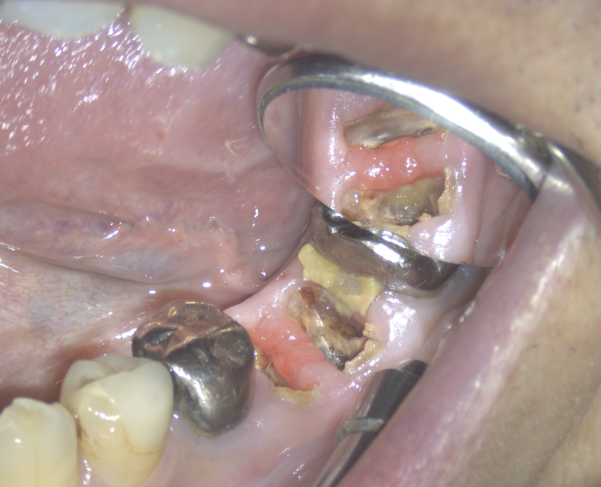
02 | Exposing the tooth covered by gum tissue and treating decay
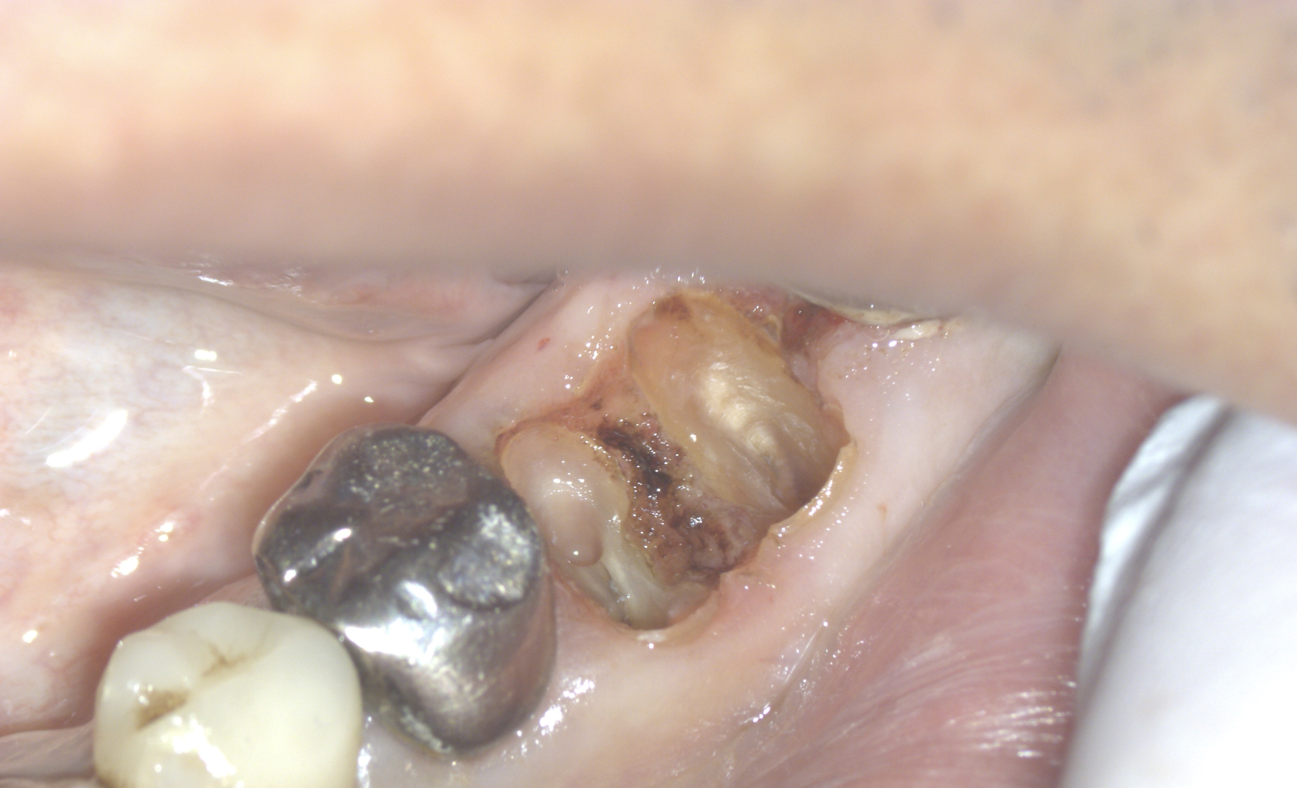
03 | Repair inside the root canals
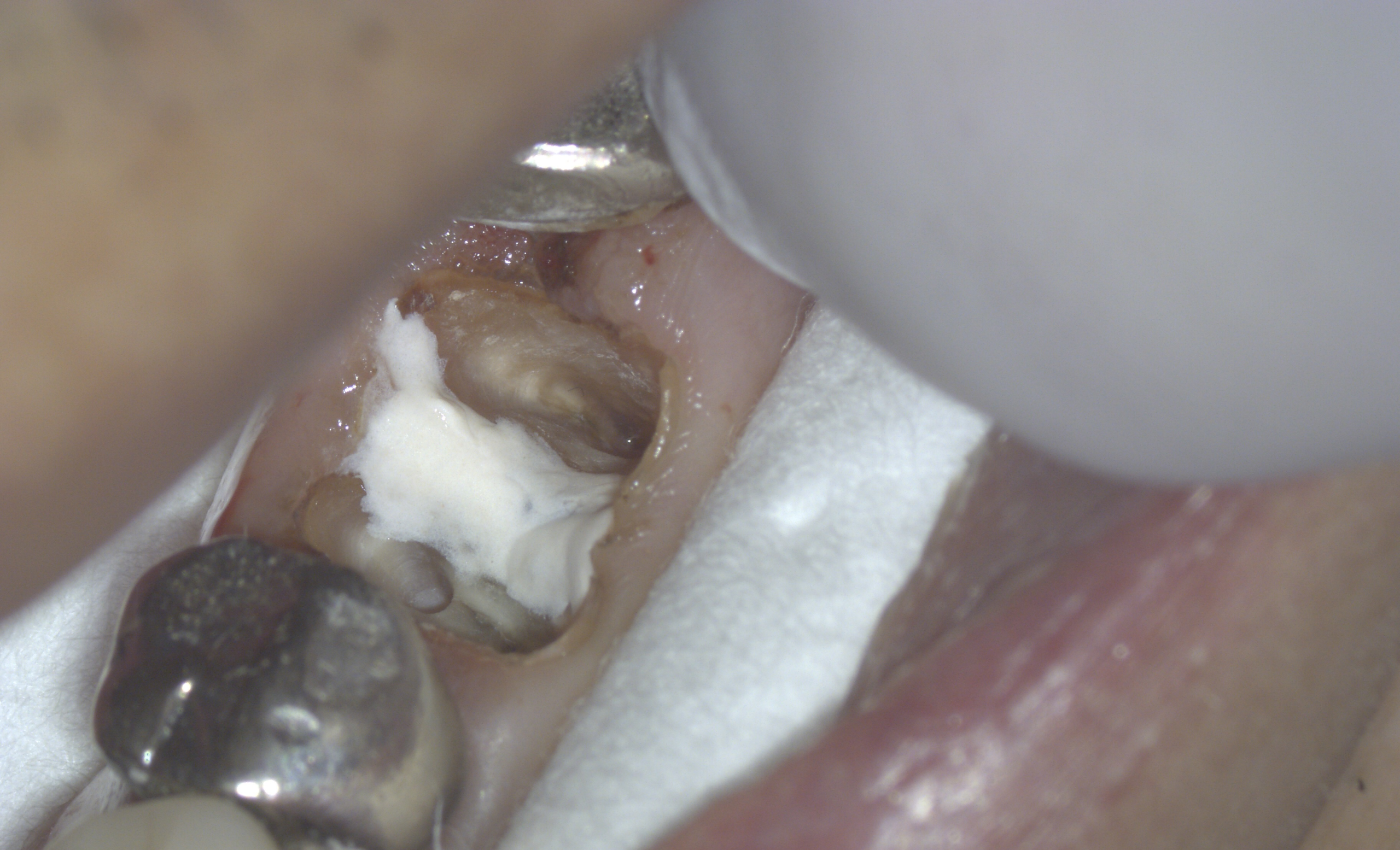
04 | Core build-up using glass-fiber posts
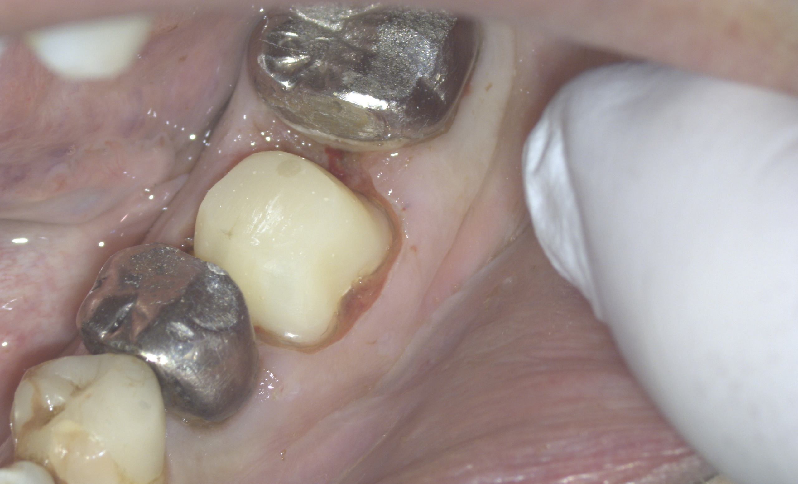
05 | Placement of a provisional crown on the same day
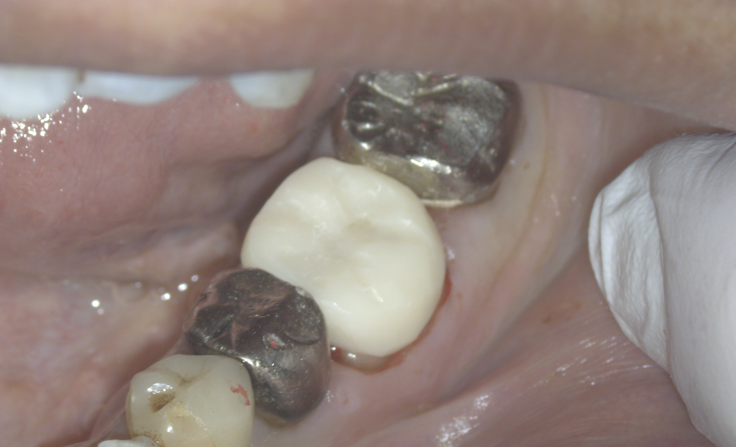
06 | Fabrication of a zirconia crown
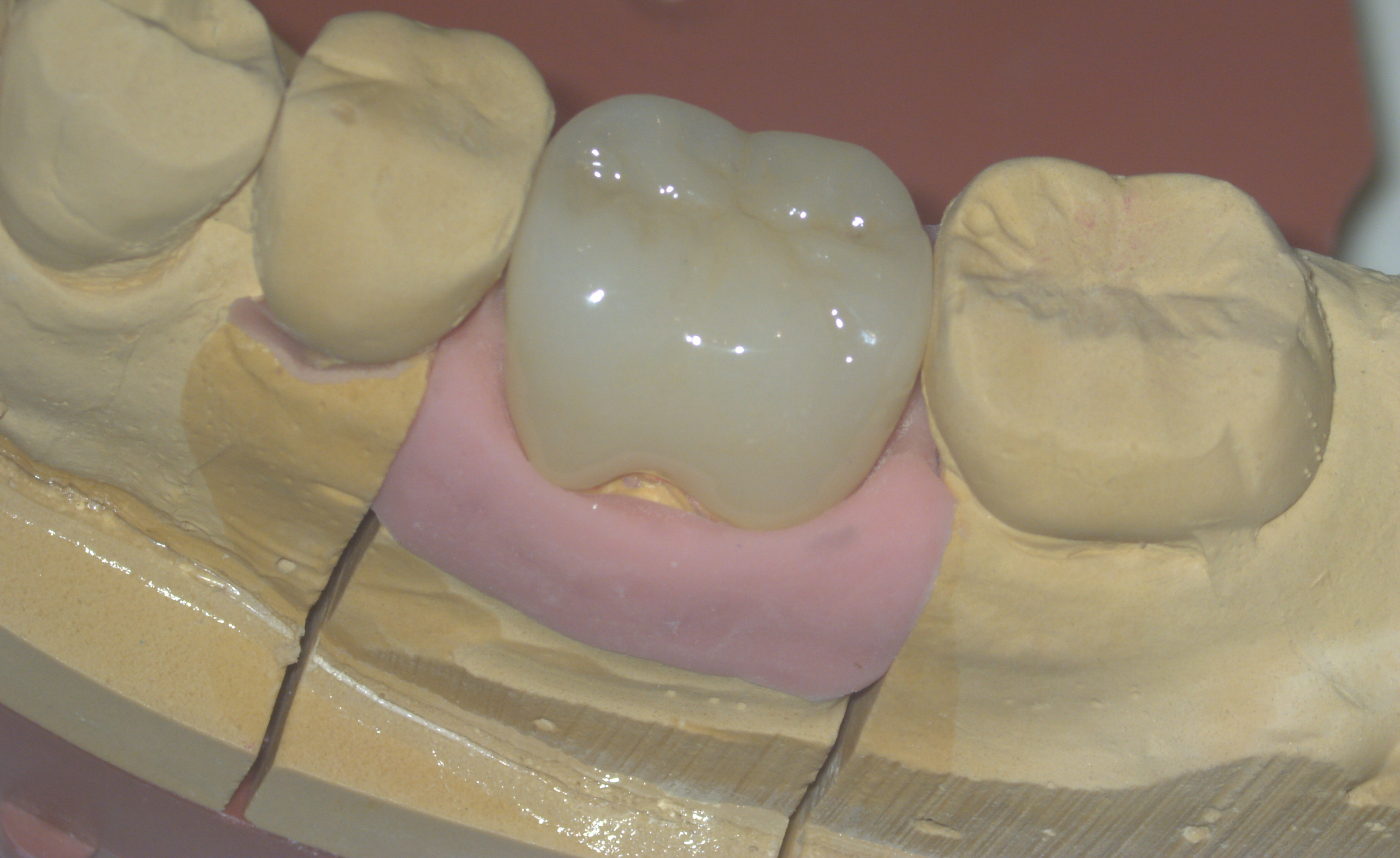
07 | Zirconia crown placement
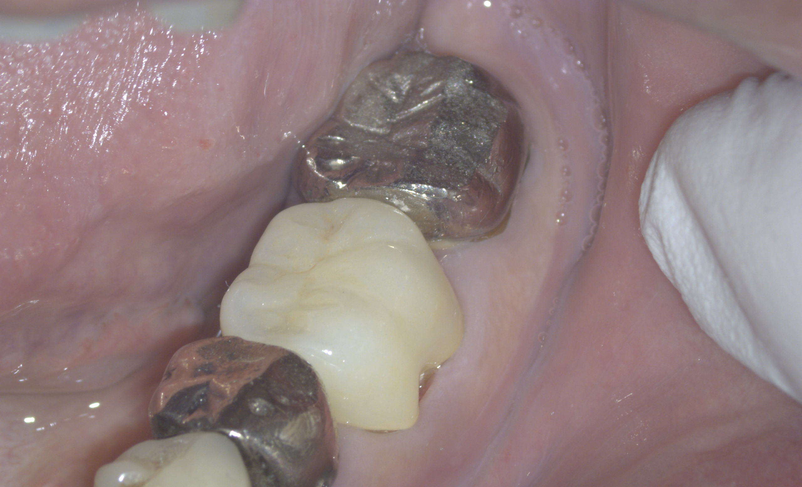
Treatment, cost, duration and number of visits for this case
Treatment: Exposure of the tooth covered by gum tissue, treatment of decay, intraoral repair inside the root canals, core build-up using glass-fiber posts, placement of a provisional crown and final restoration with a zirconia crown.
Cost: JPY 550,000 (tax included; private treatment not covered by Japanese public health insurance)
Treatment duration: Approximately 1 month
Number of visits: 3 visits
Main risks and treatment limitations
Pain, swelling or bleeding may occur after treatment. Risks include separation of the bonded area, fracture of the tooth or core, loosening or breakage of the crown, recurrent decay or infection, and gum inflammation. Further treatment or extraction may be necessary depending on the condition of the tooth and the course of treatment.
The cost, duration and number of visits refer to this case. Suitability for treatment and the course of recovery vary from person to person. This case shows the treatment sequence up to crown placement and does not guarantee preservation of the tooth or long-term treatment outcomes.
The case photographs are used with the patient’s consent to publication.