Intraoral adhesive repair of a cracked upper left molar that had been recommended for extraction
This case involved intraoral adhesive repair of a microcrack in the upper left first molar. Photographs show the treatment sequence through root canal treatment, reinforcement and zirconia crown placement.
Case video
Treatment details for this case
Treatment: The fracture line was examined under a microscope. After removing the contaminated tissue, intraoral adhesive repair was performed using Super-Bond.
Cost: JPY 550,000 (tax included)
Treatment duration: Approximately 3 months
Number of visits: 4 visits
The treatment sequence is shown from intraoral adhesive repair through root canal treatment, reinforcement with glass fiber and a resin core, and zirconia crown placement.
Main risks and treatment limitations
Reinfection, progression of the fracture or refracture may occur after treatment, and further treatment or extraction may be necessary. Outcomes vary depending on the location and extent of the fracture, the condition of the tooth and periodontal tissues, and other factors. This is one individual case and does not guarantee that every tooth can be preserved or that long-term treatment outcomes will be achieved.
Background before the first visit
During root canal treatment at the previous dental practice, a fracture line was found inside the root canal and treatment was stopped. The patient had been told that extraction was necessary. The opening photograph shows the tooth temporarily sealed with pink cement during treatment.
Rubber dam placement and examination of the fracture line
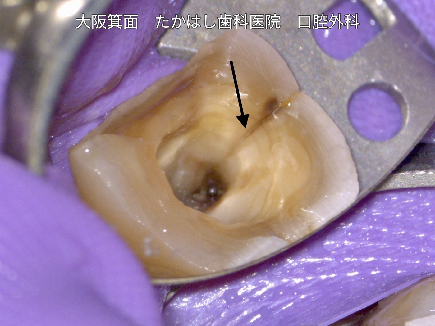
A rubber sheet called a rubber dam was placed to prevent saliva and contaminants from entering the tooth. After the temporary sealing cement was removed, a clear fracture line extending deep into the root was visible.
Removal of contaminated tissue and adhesive repair
Contamination had seeped into the fracture line and caused discoloration. Using this as a guide, the contaminated tissue was gradually removed under microscopic observation. This is a delicate procedure requiring patience.
Super-Bond was applied through the area where tissue had been removed to repair the fracture, with the aim of preventing contaminants from entering again.
From root canal treatment and reinforcement to crown placement
After disinfection and treatment inside the root canals, the canals were filled. Glass-fiber reinforcing posts and a resin core were then used to strengthen the tooth, and excess material was trimmed. Following observation, the core was shaped for the crown impression and a zirconia crown was fitted.
The case photographs and video are used with the patient’s consent to publication.