Adhesive repair and replantation of an upper right tooth with only the root remaining, split into two pieces
This case involved adhesive repair and intentional replantation of an upper right second premolar after the tooth broke, leaving only its root. The removed tooth was completely separated into two pieces. After treatment of the fracture and root, the tooth was returned to its original position. Following splinting and observation, a zirconia crown was fitted.
This is a case record from Takahashi Dental Clinic and Oral Surgery in Minoh, Osaka. The upper right second premolar is the fifth tooth from the front on the upper right.
This page contains photographs taken inside the mouth during treatment and photographs of the removed tooth.
- 01 | Before treatment: Only the root remained after the tooth broke
- 02 | Immediately after removal of the tooth
- 03 | The tooth outside the mouth
- 04 | Examination of the tooth split into two pieces
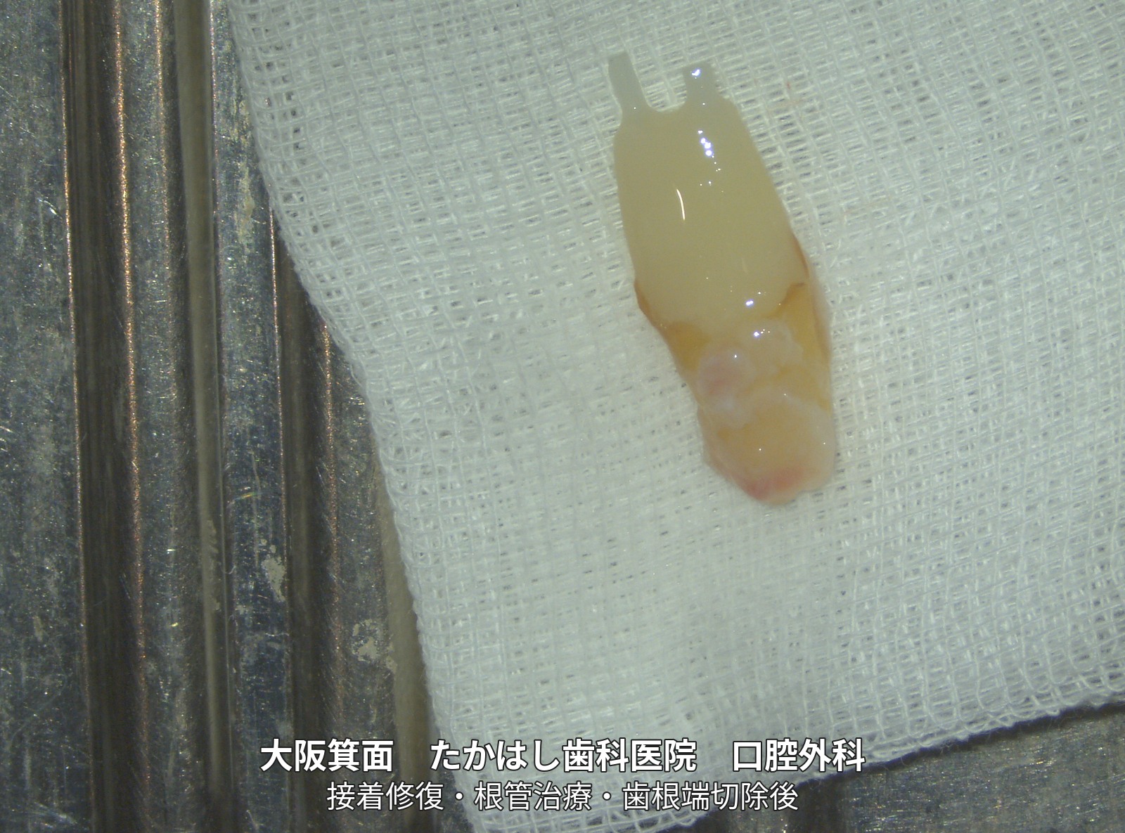
- 05 | Bonding the fracture and treating the root
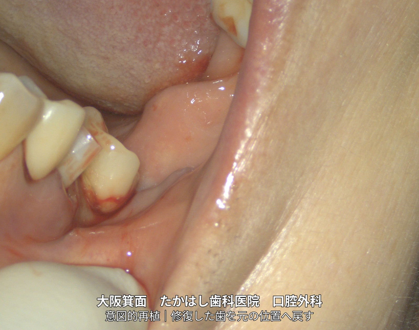
- 06 | Returning the repaired tooth to its original position
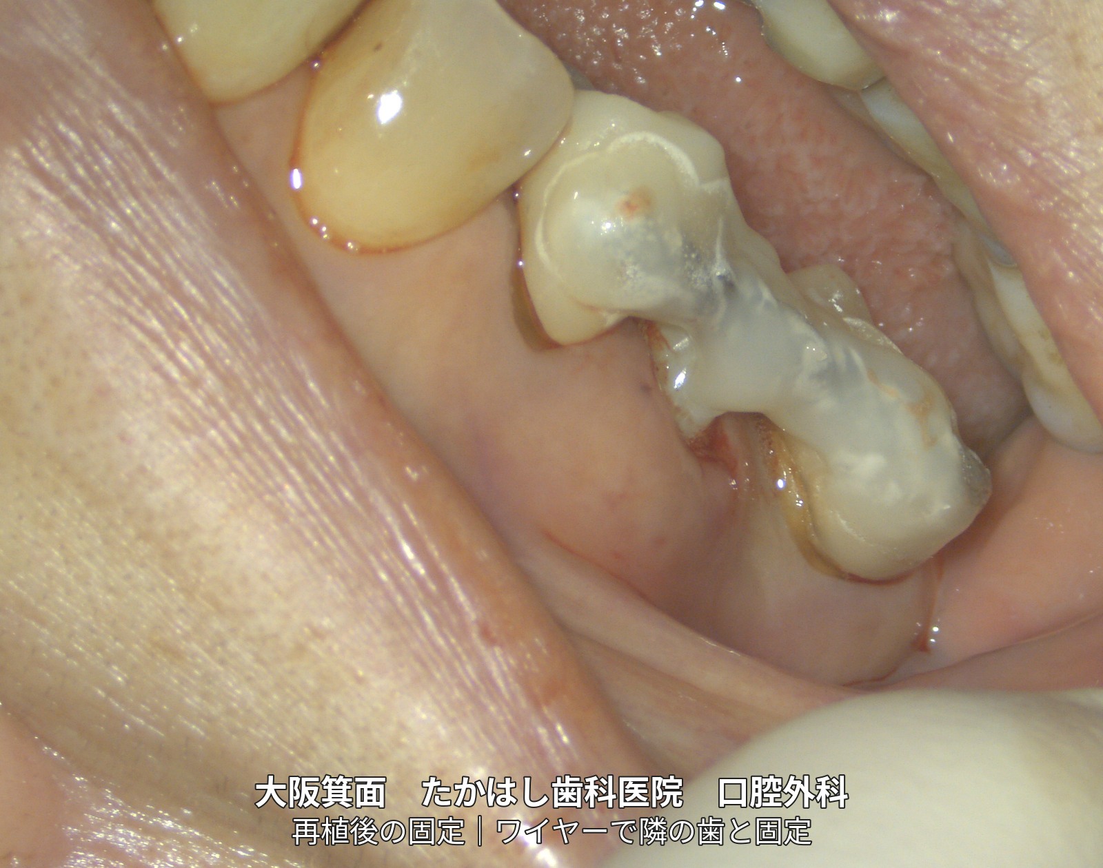
- 07 | Wire splinting to adjacent teeth
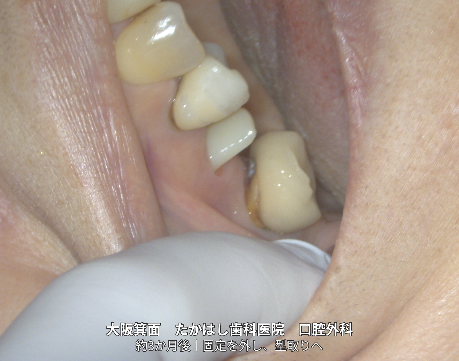
- 08 | About 3 months later: Stability checked before taking an impression
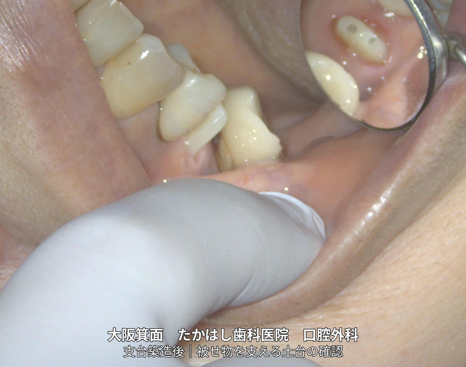
- 09 | Checking the core that supports the crown
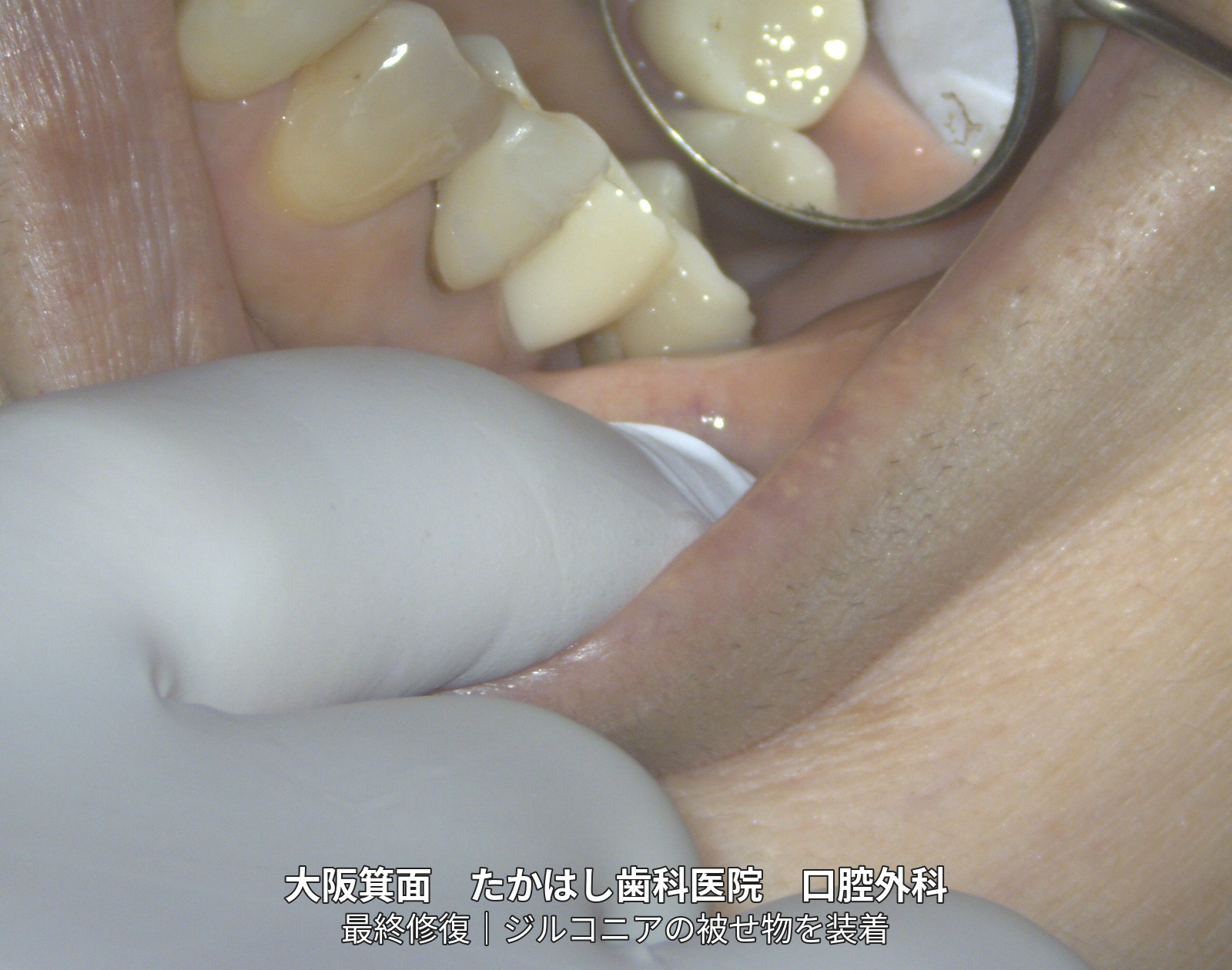
- 10 | Zirconia crown placement
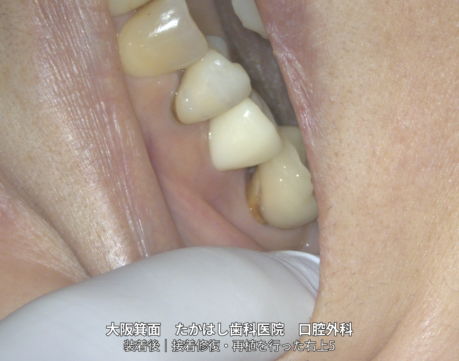
- 11 | After the final restoration: Side view
- Treatment, cost, duration and number of visits for this case
- Main risks and treatment limitations
- Related treatments and cases
01 | Before treatment: Only the root remained after the tooth broke
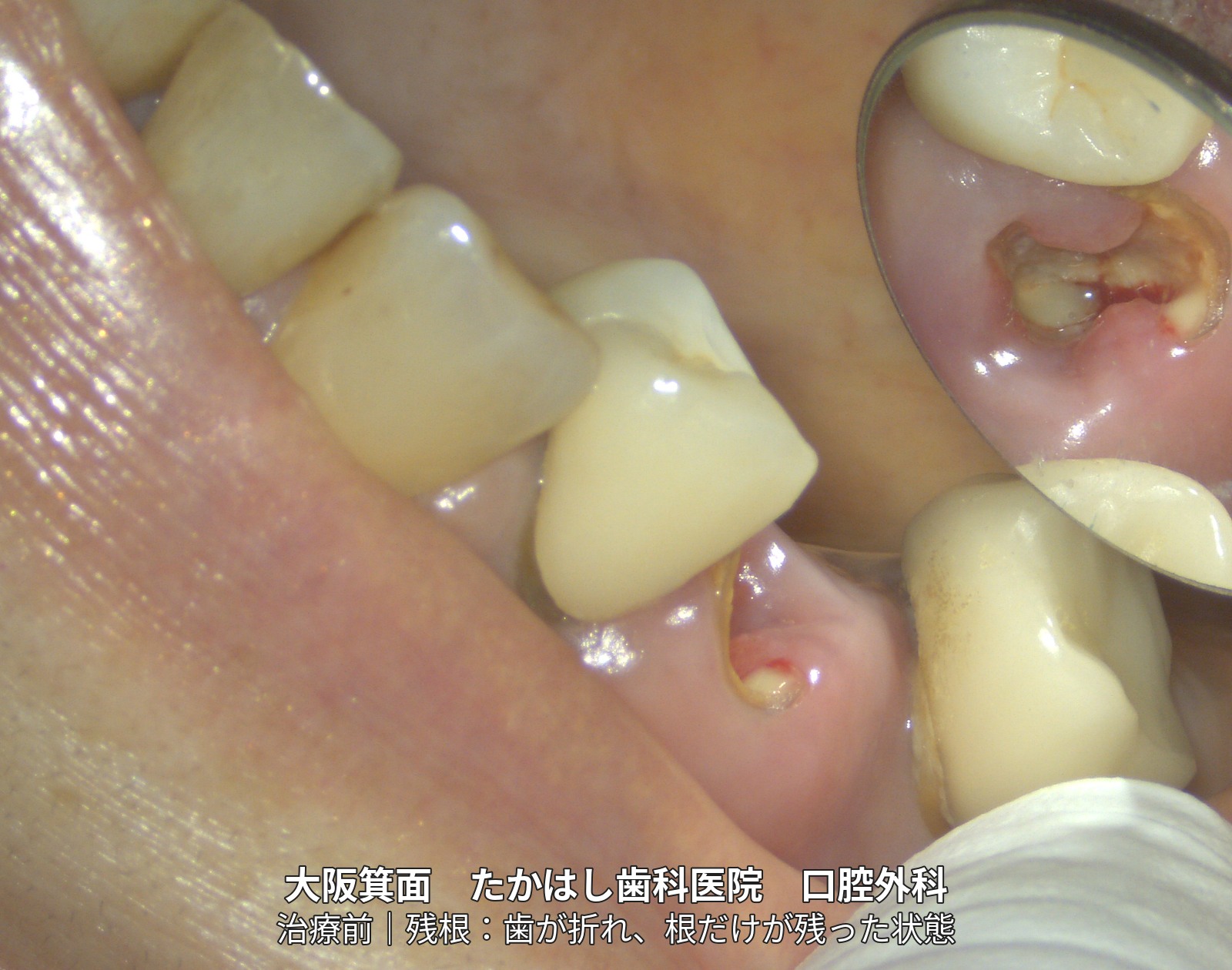
02 | Immediately after removal of the tooth
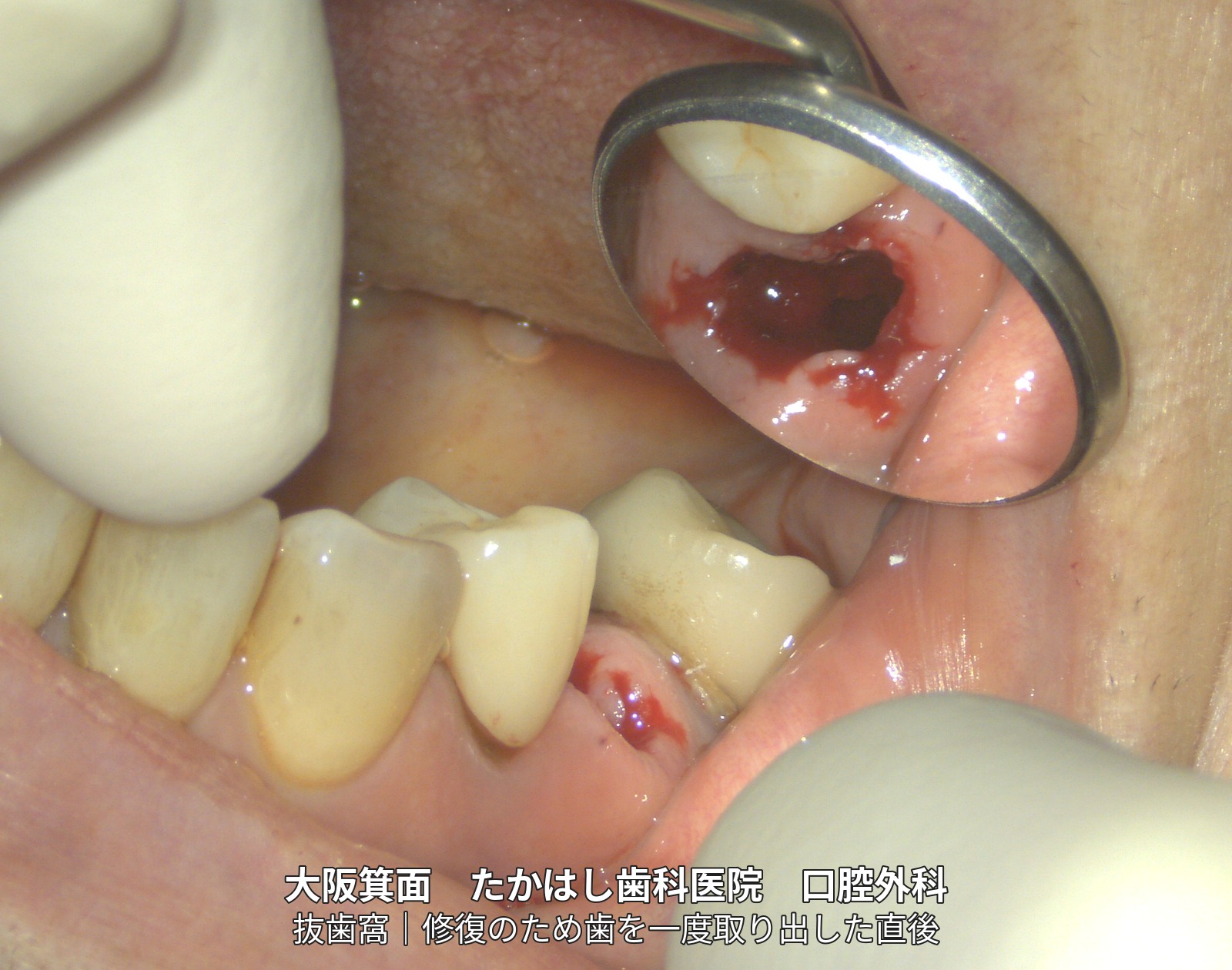
03 | The tooth outside the mouth
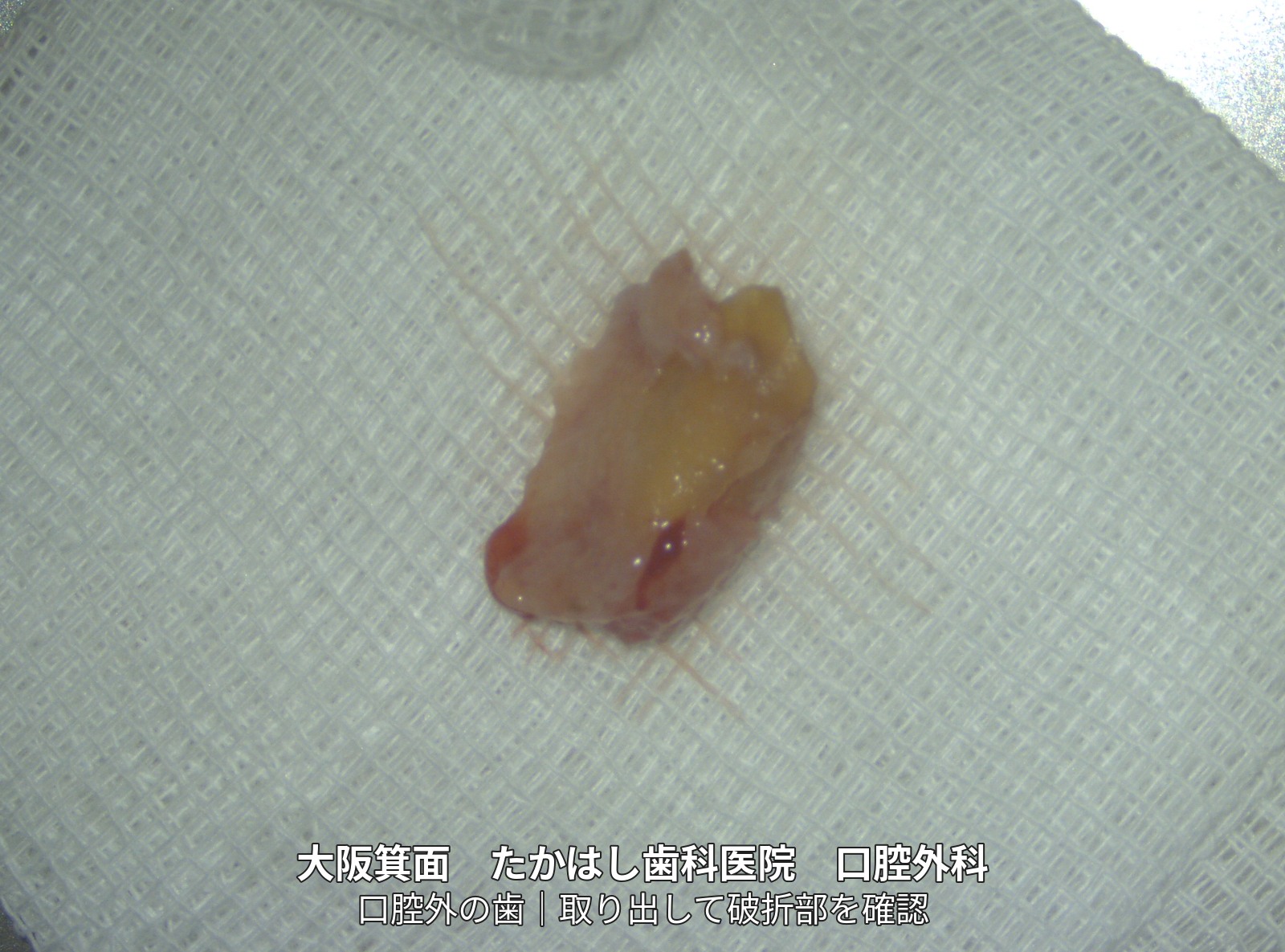
04 | Examination of the tooth split into two pieces
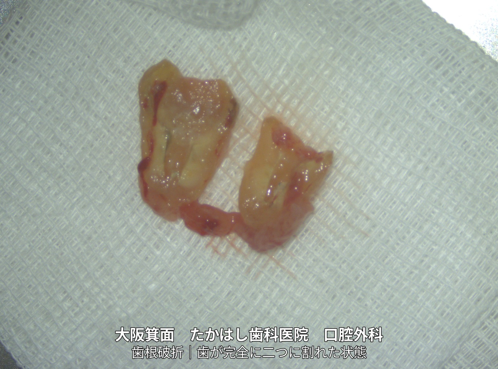
05 | Bonding the fracture and treating the root
06 | Returning the repaired tooth to its original position
07 | Wire splinting to adjacent teeth
08 | About 3 months later: Stability checked before taking an impression
09 | Checking the core that supports the crown
10 | Zirconia crown placement
11 | After the final restoration: Side view
Treatment, cost, duration and number of visits for this case
Treatment: Extraoral adhesive repair (bonding the fractured pieces together), root canal treatment (treatment inside the root), root-end resection (removal of the root tip), intentional replantation (returning the removed tooth to its original position) and splinting, core build-up (making a foundation to support the crown), and final restoration with a zirconia crown.
Total cost: JPY 640,000 (tax included, including placement of the final zirconia crown; private treatment not covered by Japanese public health insurance)
Treatment duration: Approximately 3 months through crown placement
Number of visits: 9 visits
The cost, duration and number of visits refer to this case. They vary depending on the condition of the tooth and the course of treatment.
Main risks and treatment limitations
Risks of adhesive repair and intentional replantation include refracture, infection, root resorption, ankylosis (direct fusion of the tooth to bone), and poor healing of the periodontal tissues. The tooth may not be preserved, and further treatment or extraction may be necessary. Suitability for treatment and the course of recovery vary from person to person.
This case shows the treatment sequence up to crown placement. It does not guarantee preservation of the tooth or long-term treatment outcomes.
The case photographs are used with the patient’s consent to publication.
Related treatments and cases
Adhesive repair: Bonding a fractured tooth together
Intentional replantation: Temporarily removing a tooth for treatment and returning it to its socket
Main clinic website: Tooth-preserving treatment for teeth recommended for extraction